 Explore
 Validate
 Learn
Learn Immunohistochemistry
 Flow cytometry
Antibody data
- Antibody Data
- Antigen structure
- References [17]
- Comments [0]
- Validations
- Flow cytometry [8]
Submit
Validation data
Reference
Comment
Report error
- Product number
- NBP2-25199 - Provider product page

- Provider
- Novus Biologicals
- Product name
- Mouse Monoclonal CD4 Antibody
- Antibody type
- Monoclonal
- Description
- Protein G purified.
- Reactivity
- Human
- Host
- Mouse
- Isotype
- IgG
- Vial size
- 0.1 mg
- Concentration
- 1 mg/ml
- Storage
- Store at 4C short term. Aliquot and store at -20C long term. Avoid freeze-thaw cycles.
Submitted references Glioma-derived extracellular vesicles selectively suppress immune responses.
Preferential elimination of CD28+ T cells in systemic lupus erythematosus (SLE) and the relation with activation-induced apoptosis.
Preferential elimination of CD28+ T cells in systemic lupus erythematosus (SLE) and the relation with activation-induced apoptosis.
CD40 expressed on thymic epithelial cells provides costimulation for proliferation but not for apoptosis of human thymocytes.
CD34+CD38dim cells in the human thymus can differentiate into T, natural killer, and dendritic cells but are distinct from pluripotent stem cells.
Human immunodeficiency virus type 1 gp120 induces anergy in human peripheral blood lymphocytes by inducing interleukin-10 production.
Cardiac allograft vascular disease. Relationship to microvascular cell surface markers and inflammatory cell phenotypes on endomyocardial biopsy.
Cardiac allograft vascular disease. Relationship to microvascular cell surface markers and inflammatory cell phenotypes on endomyocardial biopsy.
Development of retrovirally marked human T progenitor cells into mature thymocytes.
Development of retrovirally marked human T progenitor cells into mature thymocytes.
A monoclonal antibody (A6) recognizing a unique epitope restricted to CD45RO and RB isoforms of the leukocyte common antigen family identifies functional T cell subsets.
Screening of 78 monoclonal antibodies directed against human leukocyte antigens for cross-reactivity with surface markers on canine lymphocytes.
Screening of 78 monoclonal antibodies directed against human leukocyte antigens for cross-reactivity with surface markers on canine lymphocytes.
Phenotypic and functional analysis of T-cell precursors in the human fetal liver and thymus: CD7 expression in the early stages of T- and myeloid-cell development.
Precursors of CD3+CD4+CD8+ cells in the human thymus are defined by expression of CD34. Delineation of early events in human thymic development.
Precursors of CD3+CD4+CD8+ cells in the human thymus are defined by expression of CD34. Delineation of early events in human thymic development.
Immunofluorescent labeling using covalently linked anti-phycoerythrin antibodies and phycoerythrin polymers.
Hellwinkel JE, Redzic JS, Harland TA, Gunaydin D, Anchordoquy TJ, Graner MW
Neuro-oncology 2016 Apr;18(4):497-506
Neuro-oncology 2016 Apr;18(4):497-506
Preferential elimination of CD28+ T cells in systemic lupus erythematosus (SLE) and the relation with activation-induced apoptosis.
Kaneko H, Saito K, Hashimoto H, Yagita H, Okumura K, Azuma M
Clinical and experimental immunology 1996 Nov;106(2):218-29
Clinical and experimental immunology 1996 Nov;106(2):218-29
CD40 expressed on thymic epithelial cells provides costimulation for proliferation but not for apoptosis of human thymocytes.
Ruggiero G, Martinez Cáceres E, Voordouw A, Noteboom E, Graf D, Kroczek RA, Spits H
Journal of immunology (Baltimore, Md. : 1950) 1996 May 15;156(10):3737-46
Journal of immunology (Baltimore, Md. : 1950) 1996 May 15;156(10):3737-46
CD34+CD38dim cells in the human thymus can differentiate into T, natural killer, and dendritic cells but are distinct from pluripotent stem cells.
Res P, Martínez-Cáceres E, Cristina Jaleco A, Staal F, Noteboom E, Weijer K, Spits H
Blood 1996 Jun 15;87(12):5196-206
Blood 1996 Jun 15;87(12):5196-206
Human immunodeficiency virus type 1 gp120 induces anergy in human peripheral blood lymphocytes by inducing interleukin-10 production.
Schols D, De Clercq E
Journal of virology 1996 Aug;70(8):4953-60
Journal of virology 1996 Aug;70(8):4953-60
Cardiac allograft vascular disease. Relationship to microvascular cell surface markers and inflammatory cell phenotypes on endomyocardial biopsy.
Deng MC, Bell S, Huie P, Pinto F, Hunt SA, Stinson EB, Sibley R, Hall BM, Valantine HA
Circulation 1995 Mar 15;91(6):1647-54
Circulation 1995 Mar 15;91(6):1647-54
Development of retrovirally marked human T progenitor cells into mature thymocytes.
Staal FJ, Res PC, Weijer K, Spits H
International immunology 1995 Aug;7(8):1301-9
International immunology 1995 Aug;7(8):1301-9
Development of retrovirally marked human T progenitor cells into mature thymocytes.
Staal FJ, Res PC, Weijer K, Spits H
International immunology 1995 Aug;7(8):1301-9
International immunology 1995 Aug;7(8):1301-9
A monoclonal antibody (A6) recognizing a unique epitope restricted to CD45RO and RB isoforms of the leukocyte common antigen family identifies functional T cell subsets.
Aversa G, Waugh JA, Hall BM
Cellular immunology 1994 Oct 15;158(2):314-28
Cellular immunology 1994 Oct 15;158(2):314-28
Screening of 78 monoclonal antibodies directed against human leukocyte antigens for cross-reactivity with surface markers on canine lymphocytes.
Chabanne L, Marchal T, Kaplanski C, Fournel C, Magnol JP, Monier JC, Rigal D
Tissue antigens 1994 Mar;43(3):202-5
Tissue antigens 1994 Mar;43(3):202-5
Screening of 78 monoclonal antibodies directed against human leukocyte antigens for cross-reactivity with surface markers on canine lymphocytes.
Chabanne L, Marchal T, Kaplanski C, Fournel C, Magnol JP, Monier JC, Rigal D
Tissue antigens 1994 Mar;43(3):202-5
Tissue antigens 1994 Mar;43(3):202-5
Phenotypic and functional analysis of T-cell precursors in the human fetal liver and thymus: CD7 expression in the early stages of T- and myeloid-cell development.
Bárcena A, Muench MO, Galy AH, Cupp J, Roncarolo MG, Phillips JH, Spits H
Blood 1993 Dec 1;82(11):3401-14
Blood 1993 Dec 1;82(11):3401-14
Precursors of CD3+CD4+CD8+ cells in the human thymus are defined by expression of CD34. Delineation of early events in human thymic development.
Galy A, Verma S, Bárcena A, Spits H
The Journal of experimental medicine 1993 Aug 1;178(2):391-401
The Journal of experimental medicine 1993 Aug 1;178(2):391-401
Immunofluorescent labeling using covalently linked anti-phycoerythrin antibodies and phycoerythrin polymers.
Wilson MR, Crowley S, Odgers GA, Shaw L
Cytometry 1991;12(4):373-7
Cytometry 1991;12(4):373-7
No comments: Submit comment
Supportive validation
- Submitted by
- Novus Biologicals (provider)
- Main image
- Experimental details
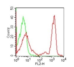
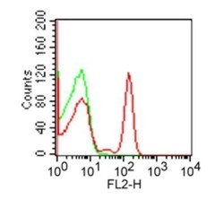
- Flow Cytometry: CD4 Antibody (RPA-T4) [NBP2-25199] - Cell surface flow analysis of CD4 in human PBMC using this antibody at 0.25 ug/10^6 cells. Cells were stained with primary antibody followed by a PE-conjugated goat anti-mouse secondary antibody this antibody . Green represents isotype control; red represents anti-CD4 antibody. Cells in the lymphocyte gate were used for analysis.
- Submitted by
- Novus Biologicals (provider)
- Main image
- Experimental details
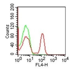
- Flow Cytometry: CD4 Antibody (RPA-T4) [NBP2-25199] - Analysis using the Allophycocyanin conjugate of NBP2-27216. Staining of CD4 in 1x10^6 human PBMC using 10 ul (0.1 ug) of was used to test this product. Propidium iodide negative lymphocyte population gated for analysis. Image using the Azide Free format of this antibody.
- Submitted by
- Novus Biologicals (provider)
- Main image
- Experimental details
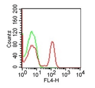
- Flow Cytometry: CD4 Antibody (RPA-T4) [NBP2-25199] - Analysis using the PE conjugate of NBP2-27216. Staining of CD4 in 1x10^6 human PBMC using 10 ul (0.1 ug) of this antibody. Green represents isotype control; red represents anti-CD4 antibody. Image using the Azide Free format of this antibody.
- Submitted by
- Novus Biologicals (provider)
- Main image

- Experimental details
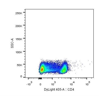
- Flow Cytometry: CD4 Antibody (RPA-T4) [NBP2-25199] - Analysis using the DyLight 405 conjugate of NBP2-27216. Staining of CD4 in human PBMCs using anti-CD4 antibody. Image from verified customer review.
- Submitted by
- Novus Biologicals (provider)
- Main image
- Experimental details
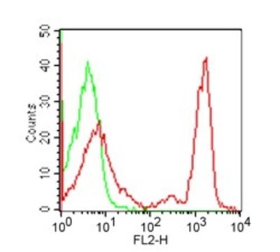
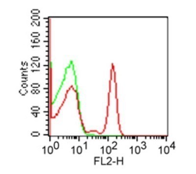
- Flow Cytometry: CD4 Antibody (RPA-T4) [NBP2-25199] - Analysis using Azide Free version of NBP2-25199. CD4 in human PBMCs using 0.1 ug of this antibody. Secondary antibody is goat anti-mouse PE.

- Submitted by
- Novus Biologicals (provider)
- Main image

- Experimental details
- Flow Cytometry: CD4 Antibody (RPA-T4) [NBP2-25199] - Analysis using the PE conjugate of NBP2-27216. Staining of CD4 in human PBMC using anti-CD4 antibody. Image from verified customer review.

- Submitted by
- Novus Biologicals (provider)
- Main image

- Experimental details
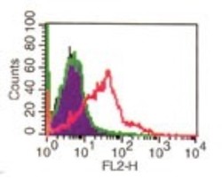
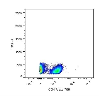
- Flow Cytometry: CD4 Antibody (RPA-T4) [NBP2-25199] - Analysis using the Alexa Fluor (R) 700 conjugate of NBP2-27216. Staining of human PBMC. Image from verified customer review.
- Submitted by
- Novus Biologicals (provider)
- Main image

- Experimental details
- Flow Cytometry: CD4 Antibody (RPA-T4) [NBP2-25199] - Analysis using the FITC conjugate of NBP2-27216. Staining of CD4 in 1x10^6 human PBMC using 10 ul (0.1 ug) of was used to test this product. Propidium iodide negative lymphocyte population gated for analysis. Image using the Azide Free format of this antibody.
