Explore
 Validate
 Learn
Learn Other assay
Antibody data
- Antibody Data
- Antigen structure
- References [7]
- Comments [0]
- Validations
- Other assay [2]
Submit
Validation data
Reference
Comment
Report error
- Product number
- MA1-7631 - Provider product page

- Provider
- Invitrogen Antibodies
- Product name
- CD4 Monoclonal Antibody (RIV6)
- Antibody type
- Monoclonal
- Antigen
- Purifed from natural sources
- Description
- MA1-7631 detects CD4 from human, rat and mouse samples.
- Antibody clone number
- RIV6
- Concentration
- 1 mg/mL
Submitted references T cell infiltration in both human multiple system atrophy and a novel mouse model of the disease.
Induction of Adaptive Immunity Leads to Nigrostriatal Disease Progression in MPTP Mouse Model of Parkinson's Disease.
In situ B cell-mediated immune responses and tubulointerstitial inflammation in human lupus nephritis.
Memory B-cell aggregates in skin biopsy are diagnostic for primary Sjögren's syndrome.
Increased intratumoral IL-17-producing cells correlate with poor survival in hepatocellular carcinoma patients.
Histopathological features in both the eschar and erythematous lesions of Tsutsugamushi Disease: identification of CD30+ cell infiltration in Tsutsugamushi disease.
BAFF-modulated repopulation of B lymphocytes in the blood and salivary glands of rituximab-treated patients with Sjögren's syndrome.
Williams GP, Marmion DJ, Schonhoff AM, Jurkuvenaite A, Won WJ, Standaert DG, Kordower JH, Harms AS
Acta neuropathologica 2020 May;139(5):855-874
Acta neuropathologica 2020 May;139(5):855-874
Induction of Adaptive Immunity Leads to Nigrostriatal Disease Progression in MPTP Mouse Model of Parkinson's Disease.
Chandra G, Roy A, Rangasamy SB, Pahan K
Journal of immunology (Baltimore, Md. : 1950) 2017 Jun 1;198(11):4312-4326
Journal of immunology (Baltimore, Md. : 1950) 2017 Jun 1;198(11):4312-4326
In situ B cell-mediated immune responses and tubulointerstitial inflammation in human lupus nephritis.
Chang A, Henderson SG, Brandt D, Liu N, Guttikonda R, Hsieh C, Kaverina N, Utset TO, Meehan SM, Quigg RJ, Meffre E, Clark MR
Journal of immunology (Baltimore, Md. : 1950) 2011 Feb 1;186(3):1849-60
Journal of immunology (Baltimore, Md. : 1950) 2011 Feb 1;186(3):1849-60
Memory B-cell aggregates in skin biopsy are diagnostic for primary Sjögren's syndrome.
Roguedas AM, Pers JO, Lemasson G, Devauchelle V, Tobón GJ, Saraux A, Misery L, Youinou P
Journal of autoimmunity 2010 Nov;35(3):241-7
Journal of autoimmunity 2010 Nov;35(3):241-7
Increased intratumoral IL-17-producing cells correlate with poor survival in hepatocellular carcinoma patients.
Zhang JP, Yan J, Xu J, Pang XH, Chen MS, Li L, Wu C, Li SP, Zheng L
Journal of hepatology 2009 May;50(5):980-9
Journal of hepatology 2009 May;50(5):980-9
Histopathological features in both the eschar and erythematous lesions of Tsutsugamushi Disease: identification of CD30+ cell infiltration in Tsutsugamushi disease.
Lee JS, Park MY, Kim YJ, Kil HI, Choi YH, Kim YC
The American Journal of dermatopathology 2009 Aug;31(6):551-6
The American Journal of dermatopathology 2009 Aug;31(6):551-6
BAFF-modulated repopulation of B lymphocytes in the blood and salivary glands of rituximab-treated patients with Sjögren's syndrome.
Pers JO, Devauchelle V, Daridon C, Bendaoud B, Le Berre R, Bordron A, Hutin P, Renaudineau Y, Dueymes M, Loisel S, Berthou C, Saraux A, Youinou P
Arthritis and rheumatism 2007 May;56(5):1464-77
Arthritis and rheumatism 2007 May;56(5):1464-77
No comments: Submit comment
Supportive validation
- Submitted by
- Invitrogen Antibodies (provider)
- Main image

- Experimental details
- NULL
- Submitted by
- Invitrogen Antibodies (provider)
- Main image

- Experimental details
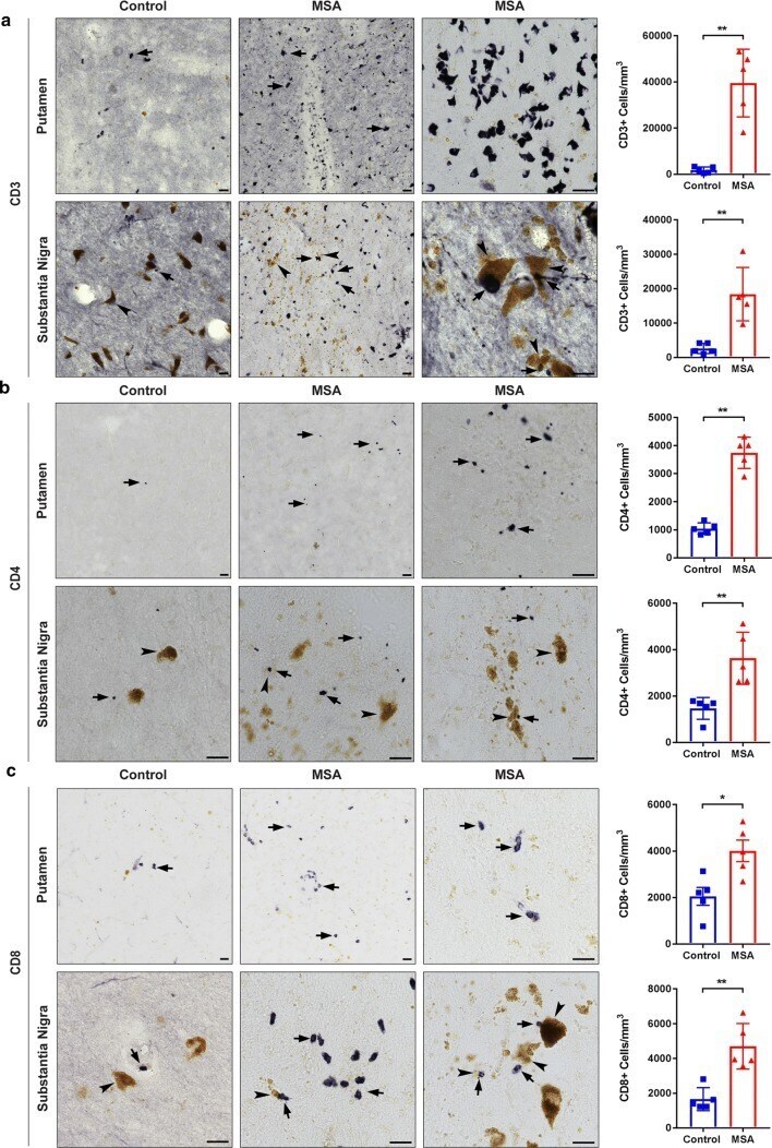
- Fig. 2 T cell infiltration in human postmortem MSA brain tissue. Control (left) and MSA (middle, right) postmortem brain sections of the putamen (top panel) and substantia nigra (bottom panel) were stained with CD3 ( a ), CD4 ( b ), and CD8 ( c ) antibodies (DAB, black staining). Unbiased stereological cell counts indicate a significant increase in the number of CD3 + T cells ( a ), CD4 + T ( b ), and CD8 + T cells ( c ) in the putamen and in the substantia nigra of MSA cases compared to control subjects. Brown coloring in the nigral sections are neuromalanin laden dopamine neurons (arrowheads). Of note, the yellow-brown pigment in putamenal sections is lipofuscin, an aging pigment composed of lipid residues of lysosomal digestion. Arrows denote T cells; arrowheads denote neuromelanin laden dopamine neurons in the substantia nigra. Representative images. All scale bars indicate 25 mum. Graphs display the mean +- SEM. * p < 0.05, ** p < 0.01, unpaired nonparametric Mann Whitney Test was used to compare groups, n = 5 per group