 Explore
 Validate
 Learn
Learn Western blot
 Immunohistochemistry
Antibody data
- Antibody Data
- Antigen structure
- References [6]
- Comments [0]
- Validations
- Immunohistochemistry [1]
- Flow cytometry [2]
- Other assay [4]
Submit
Validation data
Reference
Comment
Report error
- Product number
- 13-2444-82 - Provider product page

- Provider
- Invitrogen Antibodies
- Product name
- CD4 Monoclonal Antibody (N1UG0), Biotin, eBioscience™
- Antibody type
- Monoclonal
- Antigen
- Other
- Description
- Description: The N1UG0 monoclonal antibody reacts with human CD4, a 59 kDa cell surface glycoprotein expressed by the majority of thymocytes, a subpopulation of mature T cells (T-helper cells) and in low levels on monocytes. CD4 is a receptor for the human immunodeficiency virus (HIV). The N1UG0 antibody is recommended for use in staining human formalin-fixed paraffin embedded tissue sections. Applications Reported: This N1UG0 antibody has been reported for use in flow cytometric analysis, western blotting, and immunohistochemical staining of formalin-fixed paraffin embedded tissue sections. Applications Tested: This N1UG0 antibody has been tested by immunohistochemistry of formalin-fixed paraffin embedded human tissue using low or high pH antigen retrieval and can be used at less than or equal to 10 µg/mL. The N1UG0 antibody has been tested by flow cytometric analysis of human PBMC. This can be used at less than or equal to 0.25 µg per test. A test is defined as the amount (µg) of antibody that will stain a cell sample in a final volume of 100 µL. Cell number should be determined empirically but can range from 10^5 to 10^8 cells/test. It is recommended that the antibody be carefully titrated for optimal performance in the assay of interest. Filtration: 0.2 µm post-manufacturing filtered.
- Reactivity
- Human
- Host
- Mouse
- Conjugate
- Biotin
- Isotype
- IgG
- Antibody clone number
- N1UG0
- Vial size
- 100 µg
- Concentration
- 0.5 mg/mL
- Storage
- 4°C, store in dark, DO NOT FREEZE!
Submitted references Intrahepatic activated leukocyte cell adhesion molecule induces CD6(high)CD4(+) T cell infiltration in autoimmune hepatitis.
Asymmetric T cell division of GAD65 specific naive T cells contribute to an early divergence in the differentiation fate into memory T cell subsets.
Molecular Mechanism of Sphingosine-1-Phosphate Receptor 1 Regulating CD4(+) Tissue Memory in situ T Cells in Primary Sjogren's Syndrome.
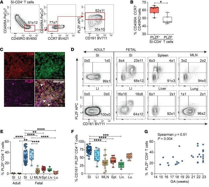
CD161 contributes to prenatal immune suppression of IFNγ-producing PLZF+ T cells.
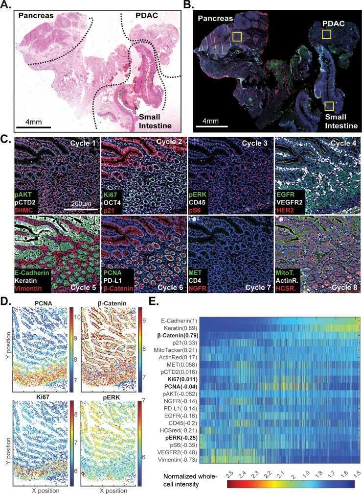
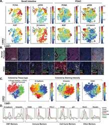
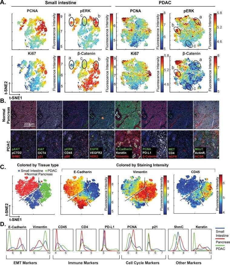
Highly multiplexed immunofluorescence imaging of human tissues and tumors using t-CyCIF and conventional optical microscopes.
Inhibition of gp160 and CD4 maturation in U937 cells after both defective and productive infections by human immunodeficiency virus type 1.
Qian Q, Cui N, Huang B, Zhao Y, Liu Q, Hu M, Li B, Wang Q, Miao Q, You Z, Ma X, Tang R
Frontiers in immunology 2022;13:967944
Frontiers in immunology 2022;13:967944
Asymmetric T cell division of GAD65 specific naive T cells contribute to an early divergence in the differentiation fate into memory T cell subsets.
Alampi G, Vignali D, Centorame I, Canu A, Cosorich I, Filoni J, Di Dedda C, Monti P
Immunology 2022 Nov;167(3):303-313
Immunology 2022 Nov;167(3):303-313
Molecular Mechanism of Sphingosine-1-Phosphate Receptor 1 Regulating CD4(+) Tissue Memory in situ T Cells in Primary Sjogren's Syndrome.
Yang XX, Yang C, Wang L, Zhou YB, Yuan X, Xiang N, Wang YP, Li XM
International journal of general medicine 2021;14:6177-6188
International journal of general medicine 2021;14:6177-6188
CD161 contributes to prenatal immune suppression of IFNγ-producing PLZF+ T cells.
Halkias J, Rackaityte E, Hillman SL, Aran D, Mendoza VF, Marshall LR, MacKenzie TC, Burt TD
The Journal of clinical investigation 2019 May 30;129(9):3562-3577
The Journal of clinical investigation 2019 May 30;129(9):3562-3577
Highly multiplexed immunofluorescence imaging of human tissues and tumors using t-CyCIF and conventional optical microscopes.
Lin JR, Izar B, Wang S, Yapp C, Mei S, Shah PM, Santagata S, Sorger PK
eLife 2018 Jul 11;7
eLife 2018 Jul 11;7
Inhibition of gp160 and CD4 maturation in U937 cells after both defective and productive infections by human immunodeficiency virus type 1.
Bour S, Boulerice F, Wainberg MA
Journal of virology 1991 Dec;65(12):6387-96
Journal of virology 1991 Dec;65(12):6387-96
No comments: Submit comment
Supportive validation
- Submitted by
- Invitrogen Antibodies (provider)
- Main image
- Experimental details
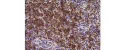
- Immunohistochemistry of formalin-fixed paraffin embedded human tonsil using 10 µg/mL of Anti-Human CD4 Biotin, followed by Streptavidin HRP, and DAB visualization. Nuclei are counterstained with hematoxylin.

Supportive validation
- Submitted by
- Invitrogen Antibodies (provider)
- Main image
- Experimental details
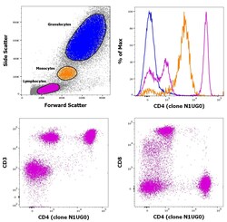
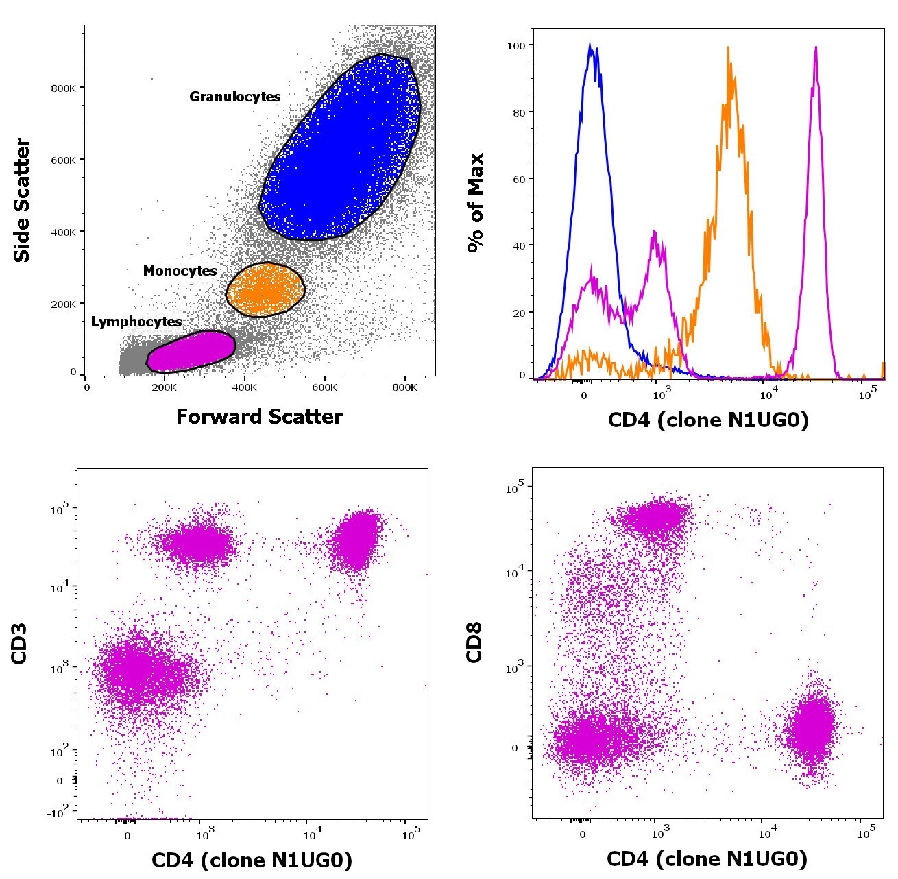
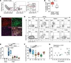
- Staining of human peripheral blood mononuclear cells with CD45 Pacific Blue, CD3 FITC, CD8 PE and CD4 Biotin followed by staining with APC conjugated streptavidin. As expected based on known relative expression patterns, CD4 clone N1UG0 stains a subset of lymphocytes (pink) and monocytes (orange), but not granulocytes (blue).
- Conjugate
- Biotin
- Submitted by
- Invitrogen Antibodies (provider)
- Main image

- Experimental details
- Staining of human peripheral blood mononuclear cells with CD45 Pacific Blue, CD3 FITC, CD8 PE and CD4 Biotin followed by staining with APC conjugated streptavidin. As expected based on known relative expression patterns, CD4 clone N1UG0 stains a subset of lymphocytes (pink) and monocytes (orange), but not granulocytes (blue).

Supportive validation
- Submitted by
- Invitrogen Antibodies (provider)
- Main image
- Experimental details
- NULL
- Conjugate
- Biotin
- Submitted by
- Invitrogen Antibodies (provider)
- Main image
- Experimental details
- NULL
- Conjugate
- Biotin
- Submitted by
- Invitrogen Antibodies (provider)
- Main image
- Experimental details
- NULL
- Conjugate
- Biotin
- Submitted by
- Invitrogen Antibodies (provider)
- Main image
- Experimental details
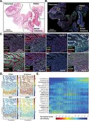
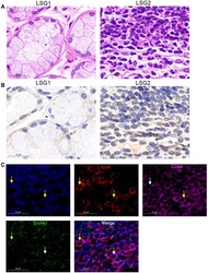
- Figure 1 S1PR1 expression in CD4 + CD69 + T RM cells. ( A ) Lymphocyte infiltration was detected by H&E staining of the labial salivary glands(LSG) of patients with pSS. ( B ) Expression of S1PR1 was detected by immunohistochemistry in labial salivary glands (LSG) of patients with pSS. ( C ) Immunofluorescence was performed to detect the expression of CD4, CD69 and S1PR1 in LSG samples with lymphocyte infiltration foci. Bar=20mum. DAPI was used to stain the nuclei and glowed blue. CD4 was pink light, CD69 was red light, and S1PR1 was green light. The yellow arrows point to positive fluorescence results and the white arrows point to negative fluorescence results in the DAPI, CD4, CD69 and S1PR1 image. In the in Merge image, the yellow arrows represent only S1PR1 expression in CD4 + T cells and no surface expression of CD69, the white arrows point to the CD4 + CD69 + T cells without the expression of S1PR1.
- Conjugate
- Biotin
