Explore
 Validate
 Learn
Learn Flow cytometry
Antibody data
- Antibody Data
- Antigen structure
- References [11]
- Comments [0]
- Validations
- Flow cytometry [1]
- Other assay [4]
Submit
Validation data
Reference
Comment
Report error
- Product number
- 47-0047-41 - Provider product page

- Provider
- Invitrogen Antibodies
- Product name
- CD4 Monoclonal Antibody (SK3 (SK-3)), APC-eFluor™ 780, eBioscience™
- Antibody type
- Monoclonal
- Antigen
- Other
- Description
- Description: The SK3 monoclonal antibody reacts with human CD4, a 59-kDa cell surface receptor expressed by a majority of thymocytes, a subpopulation of mature T helper cells, and at low levels on monocytes. CD4 is a receptor for the human immunodeficiency virus (HIV). SK3 blocks HIV binding and mixed lymphocyte reaction. The SK3 and RPA-T4 monoclonal antibodies do not cross-block binding, suggesting recognition of distinct epitopes. Applications Reported: This SK3 (SK-3) antibody has been reported for use in flow cytometric analysis. Applications Tested: This SK3 antibody has been pre-titrated and tested by flow cytometric analysis of normal human peripheral blood cells. This can be used at 5 µL (0.06 µg) per test. A test is defined as the amount (µg) of antibody that will stain a cell sample in a final volume of 100 µL. Cell number should be determined empirically but can range from 10^5 to 10^8 cells/test. APC-eFluor 780 emits at 780 nm and is excited with the Red laser (633 nm). Please make sure that your instrument is capable of detecting this fluorochome. Light sensitivity: This tandem is sensitive to photo-induced oxidation. Please protect this vial and stained samples from light. Fixation: Samples can be stored in IC Fixation Buffer (Product # 00-8222) (100 µL cell sample + 100 µL IC Fixation Buffer) or 1-step Fix/Lyse Solution (Product # 00-5333) for up to 3 days in the dark at 4°C with minimal impact on brightness and FRET efficiency/compensation. Some generalizations regarding fluorophore performance after fixation can be made, but clone specific performance should be determined empirically. Excitation: 633-647 nm; Emission: 780 nm; Laser: Red Laser. Filtration: 0.2 µm post-manufacturing filtered.
- Reactivity
- Human
- Host
- Mouse
- Isotype
- IgG
- Antibody clone number
- SK3 (SK-3)
- Vial size
- 25 Tests
- Concentration
- 5 µL/Test
- Storage
- 4° C, store in dark, DO NOT FREEZE!
Submitted references SPARC regulation of PMN clearance protects from pristane-induced lupus and rheumatoid arthritis.
CXCR3(+) T Follicular Helper Cells Induced by Co-Administration of RTS,S/AS01B and Viral-Vectored Vaccines Are Associated With Reduced Immunogenicity and Efficacy Against Malaria.
Characteristic patterns of HLA presentation and T cell differentiation in adult-onset Still's disease.
LIGHT-HVEM Signaling in Innate Lymphoid Cell Subsets Protects Against Enteric Bacterial Infection.
Tolerogenic dendritic cells generated with dexamethasone and vitamin D3 regulate rheumatoid arthritis CD4(+) T cells partly via transforming growth factor-β1.
HDAC inhibition potentiates immunotherapy in triple negative breast cancer.
IL-6-driven STAT signalling in circulating CD4+ lymphocytes is a marker for early anticitrullinated peptide antibody-negative rheumatoid arthritis.
CIB1 and CIB2 are HIV-1 helper factors involved in viral entry.
Alloantigen-specific regulatory T cells generated with a chimeric antigen receptor.
Circulating CXCR5⁺CD4⁺ T Follicular-Like Helper Cell and Memory B Cell Responses to Human Papillomavirus Vaccines.
The contribution of non-conventional T cells and NK cells in the mycobacterial-specific IFNγ response in Bacille Calmette-Guérin (BCG)-immunized infants.
Sangaletti S, Botti L, Gulino A, Lecis D, Bassani B, Portararo P, Milani M, Cancila V, De Cecco L, Dugo M, Tripodo C, Colombo MP
iScience 2021 Jun 25;24(6):102510
iScience 2021 Jun 25;24(6):102510
CXCR3(+) T Follicular Helper Cells Induced by Co-Administration of RTS,S/AS01B and Viral-Vectored Vaccines Are Associated With Reduced Immunogenicity and Efficacy Against Malaria.
Bowyer G, Grobbelaar A, Rampling T, Venkatraman N, Morelle D, Ballou RW, Hill AVS, Ewer KJ
Frontiers in immunology 2018;9:1660
Frontiers in immunology 2018;9:1660
Characteristic patterns of HLA presentation and T cell differentiation in adult-onset Still's disease.
Jung JY, Choi B, Sayeed HM, Suh CH, Kim YW, Kim HA, Sohn S
International journal of immunopathology and pharmacology 2018 Jan-Dec;32:2058738418791284
International journal of immunopathology and pharmacology 2018 Jan-Dec;32:2058738418791284
LIGHT-HVEM Signaling in Innate Lymphoid Cell Subsets Protects Against Enteric Bacterial Infection.
Seo GY, Shui JW, Takahashi D, Song C, Wang Q, Kim K, Mikulski Z, Chandra S, Giles DA, Zahner S, Kim PH, Cheroutre H, Colonna M, Kronenberg M
Cell host & microbe 2018 Aug 8;24(2):249-260.e4
Cell host & microbe 2018 Aug 8;24(2):249-260.e4
Tolerogenic dendritic cells generated with dexamethasone and vitamin D3 regulate rheumatoid arthritis CD4(+) T cells partly via transforming growth factor-β1.
Anderson AE, Swan DJ, Wong OY, Buck M, Eltherington O, Harry RA, Patterson AM, Pratt AG, Reynolds G, Doran JP, Kirby JA, Isaacs JD, Hilkens CM
Clinical and experimental immunology 2017 Jan;187(1):113-123
Clinical and experimental immunology 2017 Jan;187(1):113-123
HDAC inhibition potentiates immunotherapy in triple negative breast cancer.
Terranova-Barberio M, Thomas S, Ali N, Pawlowska N, Park J, Krings G, Rosenblum MD, Budillon A, Munster PN
Oncotarget 2017 Dec 26;8(69):114156-114172
Oncotarget 2017 Dec 26;8(69):114156-114172
IL-6-driven STAT signalling in circulating CD4+ lymphocytes is a marker for early anticitrullinated peptide antibody-negative rheumatoid arthritis.
Anderson AE, Pratt AG, Sedhom MA, Doran JP, Routledge C, Hargreaves B, Brown PM, Lê Cao KA, Isaacs JD, Thomas R
Annals of the rheumatic diseases 2016 Feb;75(2):466-73
Annals of the rheumatic diseases 2016 Feb;75(2):466-73
CIB1 and CIB2 are HIV-1 helper factors involved in viral entry.
Godinho-Santos A, Hance AJ, Gonçalves J, Mammano F
Scientific reports 2016 Aug 4;6:30927
Scientific reports 2016 Aug 4;6:30927
Alloantigen-specific regulatory T cells generated with a chimeric antigen receptor.
MacDonald KG, Hoeppli RE, Huang Q, Gillies J, Luciani DS, Orban PC, Broady R, Levings MK
The Journal of clinical investigation 2016 Apr 1;126(4):1413-24
The Journal of clinical investigation 2016 Apr 1;126(4):1413-24
Circulating CXCR5⁺CD4⁺ T Follicular-Like Helper Cell and Memory B Cell Responses to Human Papillomavirus Vaccines.
Matsui K, Adelsberger JW, Kemp TJ, Baseler MW, Ledgerwood JE, Pinto LA
PloS one 2015;10(9):e0137195
PloS one 2015;10(9):e0137195
The contribution of non-conventional T cells and NK cells in the mycobacterial-specific IFNγ response in Bacille Calmette-Guérin (BCG)-immunized infants.
Zufferey C, Germano S, Dutta B, Ritz N, Curtis N
PloS one 2013;8(10):e77334
PloS one 2013;8(10):e77334
No comments: Submit comment
Supportive validation
- Submitted by
- Invitrogen Antibodies (provider)
- Main image

- Experimental details
- Staining of normal human peripheral blood cells with Anti-Human CD8a APC (Product # 17-0088-42) and Mouse IgG1 K Isotype Control APC-eFluor® 780 (Product # 47-4714-82) (left) or Anti-Human CD4 APC-eFluor® 780 (right). Cells in the lymphocyte gate were used for analysis.

Supportive validation
- Submitted by
- Invitrogen Antibodies (provider)
- Main image
- Experimental details
- NULL

- Submitted by
- Invitrogen Antibodies (provider)
- Main image

- Experimental details
- NULL
- Submitted by
- Invitrogen Antibodies (provider)
- Main image
- Experimental details
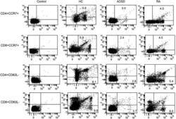
- Supplement Figure 2. Representative examples of flow cytometric dot plots of surface-stained cells presenting CD4+CCR7+, CD8+CCR7+, CD4+CD62L-, and CD8+CD62L- from the peripheral blood of one patient with adult-onset Still's disease (AOSD), a patient with rheumatoid arthritis (RA), and a healthy control (HC).

- Submitted by
- Invitrogen Antibodies (provider)
- Main image

- Experimental details
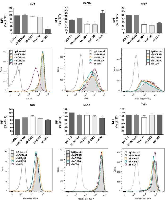
- Figure 9 Effect of downmodulation of CD4 and CIB proteins in CD4+ T-lymphocytes on the expression of proteins implicated in HIV-1 entry. Activated CD4+ T-lymphocytes were transduced with the empty vector (pLKO.1) or vectors leading to the expression of the indicated shRNAs, and the surface expression of the following molecules was measured by flow cytometry: CD3, CD4, CXCR4, LFA-1 and alpha4beta7. Intracellular talin expression was also evaluated in permeabilized cells. In each case, the mean fluorescence intensity (MFI) and a representative fluorescence histogram are shown. For MFI, the values represent the mean +- SEM of 4 independent experiments using two different shRNAs per targeted gene and are expressed relative to values obtained for non-transduced cells. *P < 0.05, **P < 0.01 versus sh-SCRAM (Kruskal-Wallis test).