 Explore
 Validate
 Learn
Learn Flow cytometry
Antibody data
- Antibody Data
- Antigen structure
- References [15]
- Comments [0]
- Validations
- Flow cytometry [1]
Submit
Validation data
Reference
Comment
Report error
- Product number
- 47-0048-41 - Provider product page

- Provider
- Invitrogen Antibodies
- Product name
- Anti-CD4 Monoclonal Antibody (OKT4 (OKT-4)), APC-eFluor 780, eBioscience™
- Antibody type
- Monoclonal
- Antigen
- Other
- Description
- Description: The OKT4 monoclonal antibody reacts with human CD4, a 59 kDa cell surface glycoprotein expressed by the majority of thymocytes, a subpopulation of mature T cells (T-helper cells) and in low levels on monocytes. CD4 is a receptor for the human immunodeficiency virus (HIV). The OKT4 antibody recognizes a different epitope than the RPA-T4 monoclonal antibody, and these antibodies do not cross-block binding to each other's respective epitopes. Applications Reported: This OKT4 (OKT-4) antibody has been reported for use in flow cytometric analysis. Applications Tested: This OKT4 (OKT-4) antibody has been pre-titrated and tested by flow cytometric analysis of normal human peripheral blood cells. This can be used at 5 µL (0.125 µg) per test. A test is defined as the amount (µg) of antibody that will stain a cell sample in a final volume of 100 µL. Cell number should be determined empirically but can range from 10^5 to 10^8 cells/test. APC-eFluor 780 emits at 780 nm and is excited with the Red laser (633 nm). Please make sure that your instrument is capable of detecting this fluorochome. Light sensitivity: This tandem is sensitive to photo-induced oxidation. Please protect this vial and stained samples from light. Fixation: Samples can be stored in IC Fixation Buffer (cat. 00-8222) (100 µL cell sample + 100 µL IC Fixation Buffer) or 1-step Fix/Lyse Solution (cat. 00-5333) for up to 3 days in the dark at 4°C with minimal impact on brightness and FRET efficiency/compensation. Some generalizations regarding fluorophore performance after fixation can be made, but clone specific performance should be determined empirically. Excitation: 633-647 nm; Emission: 780 nm; Laser: Red Laser. Filtration: 0.2 µm post-manufacturing filtered.
- Reactivity
- Human
- Host
- Mouse
- Isotype
- IgG
- Antibody clone number
- OKT4 (OKT-4)
- Vial size
- 25 Tests
- Concentration
- 5 µL/Test
- Storage
- 4° C, store in dark, DO NOT FREEZE!
Submitted references Expression of CD20 after viral reactivation renders HIV-reservoir cells susceptible to Rituximab.
Complement receptor CD46 co-stimulates optimal human CD8+ T cell effector function via fatty acid metabolism.
Accumulation of T-helper 22 cells, interleukin-22 and myeloid-derived suppressor cells promotes gastric cancer progression in elderly patients.
IL-6 receptor blockade corrects defects of XIAP-deficient regulatory T cells.
Targeted Disruption of TCF12 Reveals HEB as Essential in Human Mesodermal Specification and Hematopoiesis.
Ex vivo culture of human atherosclerotic plaques: A model to study immune cells in atherogenesis.
Single-cell profiling reveals GPCR heterogeneity and functional patterning during neuroinflammation.
Tax and Semaphorin 4D Released from Lymphocytes Infected with Human Lymphotropic Virus Type 1 and Their Effect on Neurite Growth.
PD-L1 expression and prognostic impact in glioblastoma.
Tumor-derived exosomes regulate expression of immune function-related genes in human T cell subsets.
The HIV-1 gp120 CD4-bound conformation is preferentially targeted by antibody-dependent cellular cytotoxicity-mediating antibodies in sera from HIV-1-infected individuals.
Aging and cytomegalovirus infection differentially and jointly affect distinct circulating T cell subsets in humans.
CD4-CCR5 interaction in intracellular compartments contributes to receptor expression at the cell surface.
Inhibition of gp160 and CD4 maturation in U937 cells after both defective and productive infections by human immunodeficiency virus type 1.
Separation of functional subsets of human T cells by a monoclonal antibody.
Serra-Peinado C, Grau-Expósito J, Luque-Ballesteros L, Astorga-Gamaza A, Navarro J, Gallego-Rodriguez J, Martin M, Curran A, Burgos J, Ribera E, Raventós B, Willekens R, Torrella A, Planas B, Badía R, Garcia F, Castellví J, Genescà M, Falcó V, Buzon MJ
Nature communications 2019 Aug 16;10(1):3705
Nature communications 2019 Aug 16;10(1):3705
Complement receptor CD46 co-stimulates optimal human CD8+ T cell effector function via fatty acid metabolism.
Arbore G, West EE, Rahman J, Le Friec G, Niyonzima N, Pirooznia M, Tunc I, Pavlidis P, Powell N, Li Y, Liu P, Servais A, Couzi L, Fremeaux-Bacchi V, Placais L, Ferraro A, Walsh PR, Kavanagh D, Afzali B, Lavender P, Lachmann HJ, Kemper C
Nature communications 2018 Oct 10;9(1):4186
Nature communications 2018 Oct 10;9(1):4186
Accumulation of T-helper 22 cells, interleukin-22 and myeloid-derived suppressor cells promotes gastric cancer progression in elderly patients.
Chen X, Wang Y, Wang J, Wen J, Jia X, Wang X, Zhang H
Oncology letters 2018 Jul;16(1):253-261
Oncology letters 2018 Jul;16(1):253-261
IL-6 receptor blockade corrects defects of XIAP-deficient regulatory T cells.
Hsieh WC, Hsu TS, Chang YJ, Lai MZ
Nature communications 2018 Jan 31;9(1):463
Nature communications 2018 Jan 31;9(1):463
Targeted Disruption of TCF12 Reveals HEB as Essential in Human Mesodermal Specification and Hematopoiesis.
Li Y, Brauer PM, Singh J, Xhiku S, Yoganathan K, Zúñiga-Pflücker JC, Anderson MK
Stem cell reports 2017 Sep 12;9(3):779-795
Stem cell reports 2017 Sep 12;9(3):779-795
Ex vivo culture of human atherosclerotic plaques: A model to study immune cells in atherogenesis.
Lebedeva A, Vorobyeva D, Vagida M, Ivanova O, Felker E, Fitzgerald W, Danilova N, Gontarenko V, Shpektor A, Vasilieva E, Margolis L
Atherosclerosis 2017 Dec;267:90-98
Atherosclerosis 2017 Dec;267:90-98
Single-cell profiling reveals GPCR heterogeneity and functional patterning during neuroinflammation.
Tischner D, Grimm M, Kaur H, Staudenraus D, Carvalho J, Looso M, Günther S, Wanke F, Moos S, Siller N, Breuer J, Schwab N, Zipp F, Waisman A, Kurschus FC, Offermanns S, Wettschureck N
JCI insight 2017 Aug 3;2(15)
JCI insight 2017 Aug 3;2(15)
Tax and Semaphorin 4D Released from Lymphocytes Infected with Human Lymphotropic Virus Type 1 and Their Effect on Neurite Growth.
Quintremil S, Alberti C, Rivera M, Medina F, Puente J, Cartier L, Ramírez E, Tanaka Y, Valenzuela MA
AIDS research and human retroviruses 2016 Jan;32(1):68-79
AIDS research and human retroviruses 2016 Jan;32(1):68-79
PD-L1 expression and prognostic impact in glioblastoma.
Nduom EK, Wei J, Yaghi NK, Huang N, Kong LY, Gabrusiewicz K, Ling X, Zhou S, Ivan C, Chen JQ, Burks JK, Fuller GN, Calin GA, Conrad CA, Creasy C, Ritthipichai K, Radvanyi L, Heimberger AB
Neuro-oncology 2016 Feb;18(2):195-205
Neuro-oncology 2016 Feb;18(2):195-205
Tumor-derived exosomes regulate expression of immune function-related genes in human T cell subsets.
Muller L, Mitsuhashi M, Simms P, Gooding WE, Whiteside TL
Scientific reports 2016 Feb 4;6:20254
Scientific reports 2016 Feb 4;6:20254
The HIV-1 gp120 CD4-bound conformation is preferentially targeted by antibody-dependent cellular cytotoxicity-mediating antibodies in sera from HIV-1-infected individuals.
Veillette M, Coutu M, Richard J, Batraville LA, Dagher O, Bernard N, Tremblay C, Kaufmann DE, Roger M, Finzi A
Journal of virology 2015 Jan;89(1):545-51
Journal of virology 2015 Jan;89(1):545-51
Aging and cytomegalovirus infection differentially and jointly affect distinct circulating T cell subsets in humans.
Wertheimer AM, Bennett MS, Park B, Uhrlaub JL, Martinez C, Pulko V, Currier NL, Nikolich-Žugich D, Kaye J, Nikolich-Žugich J
Journal of immunology (Baltimore, Md. : 1950) 2014 Mar 1;192(5):2143-55
Journal of immunology (Baltimore, Md. : 1950) 2014 Mar 1;192(5):2143-55
CD4-CCR5 interaction in intracellular compartments contributes to receptor expression at the cell surface.
Achour L, Scott MG, Shirvani H, Thuret A, Bismuth G, Labbé-Jullié C, Marullo S
Blood 2009 Feb 26;113(9):1938-47
Blood 2009 Feb 26;113(9):1938-47
Inhibition of gp160 and CD4 maturation in U937 cells after both defective and productive infections by human immunodeficiency virus type 1.
Bour S, Boulerice F, Wainberg MA
Journal of virology 1991 Dec;65(12):6387-96
Journal of virology 1991 Dec;65(12):6387-96
Separation of functional subsets of human T cells by a monoclonal antibody.
Reinherz EL, Kung PC, Goldstein G, Schlossman SF
Proceedings of the National Academy of Sciences of the United States of America 1979 Aug;76(8):4061-5
Proceedings of the National Academy of Sciences of the United States of America 1979 Aug;76(8):4061-5
No comments: Submit comment
Supportive validation
- Submitted by
- Invitrogen Antibodies (provider)
- Main image
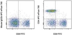
- Experimental details
- Staining of normal human peripheral blood cells with Anti-Human CD8a FITC (Product # 11-0088-42) and Mouse IgG2b K Isotype Control APC-eFluor® 780 (Product # 47-4732-80) (left) or Anti-Human CD4 APC-eFluor® 780 (right). Cells in the lymphocyte gate were used for analysis.
