Explore
 Validate
 Learn
Learn Flow cytometry
Antibody data
- Antibody Data
- Antigen structure
- References [4]
- Comments [0]
- Validations
- Flow cytometry [1]
- Other assay [4]
Submit
Validation data
Reference
Comment
Report error
- Product number
- 62-0047-42 - Provider product page

- Provider
- Invitrogen Antibodies
- Product name
- CD4 Monoclonal Antibody (SK3 (SK-3)), Super Bright™ 436, eBioscience™
- Antibody type
- Monoclonal
- Antigen
- Other
- Description
- Description: The SK3 monoclonal antibody reacts with human CD4, a 59-kDa cell surface receptor expressed by a majority of thymocytes, a subpopulation of mature T helper cells, and at low levels on monocytes. CD4 is a receptor for the human immunodeficiency virus (HIV). SK3 blocks HIV binding and mixed lymphocyte reaction. The SK3 and RPA-T4 monoclonal antibodies do not cross-block binding, suggesting recognition of distinct epitopes.
- Antibody clone number
- SK3 (SK-3)
- Concentration
- 5 µL/Test
Submitted references Characteristic patterns of HLA presentation and T cell differentiation in adult-onset Still's disease.
HDAC inhibition potentiates immunotherapy in triple negative breast cancer.
CIB1 and CIB2 are HIV-1 helper factors involved in viral entry.
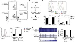
Alloantigen-specific regulatory T cells generated with a chimeric antigen receptor.
Jung JY, Choi B, Sayeed HM, Suh CH, Kim YW, Kim HA, Sohn S
International journal of immunopathology and pharmacology 2018 Jan-Dec;32:2058738418791284
International journal of immunopathology and pharmacology 2018 Jan-Dec;32:2058738418791284
HDAC inhibition potentiates immunotherapy in triple negative breast cancer.
Terranova-Barberio M, Thomas S, Ali N, Pawlowska N, Park J, Krings G, Rosenblum MD, Budillon A, Munster PN
Oncotarget 2017 Dec 26;8(69):114156-114172
Oncotarget 2017 Dec 26;8(69):114156-114172
CIB1 and CIB2 are HIV-1 helper factors involved in viral entry.
Godinho-Santos A, Hance AJ, Gonçalves J, Mammano F
Scientific reports 2016 Aug 4;6:30927
Scientific reports 2016 Aug 4;6:30927
Alloantigen-specific regulatory T cells generated with a chimeric antigen receptor.
MacDonald KG, Hoeppli RE, Huang Q, Gillies J, Luciani DS, Orban PC, Broady R, Levings MK
The Journal of clinical investigation 2016 Apr 1;126(4):1413-24
The Journal of clinical investigation 2016 Apr 1;126(4):1413-24
No comments: Submit comment
Supportive validation
- Submitted by
- Invitrogen Antibodies (provider)
- Main image

- Experimental details
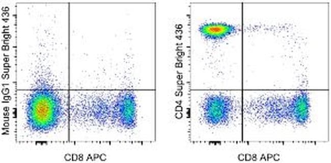
- Staining of normal human peripheral blood cells with Anti-Human CD8a APC (Product # 17-0088-42) and Mouse IgG1 K Isotype Control Super Bright 436 (Product # 62-4714-82) (left) or Anti-Human CD4 Super Bright 436 (right). Cells in the lymphocyte gate were used for analysis.
Supportive validation
- Submitted by
- Invitrogen Antibodies (provider)
- Main image
- Experimental details
- NULL

- Submitted by
- Invitrogen Antibodies (provider)
- Main image

- Experimental details
- NULL
- Submitted by
- Invitrogen Antibodies (provider)
- Main image
- Experimental details
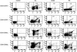
- Supplement Figure 2. Representative examples of flow cytometric dot plots of surface-stained cells presenting CD4+CCR7+, CD8+CCR7+, CD4+CD62L-, and CD8+CD62L- from the peripheral blood of one patient with adult-onset Still's disease (AOSD), a patient with rheumatoid arthritis (RA), and a healthy control (HC).

- Submitted by
- Invitrogen Antibodies (provider)
- Main image

- Experimental details
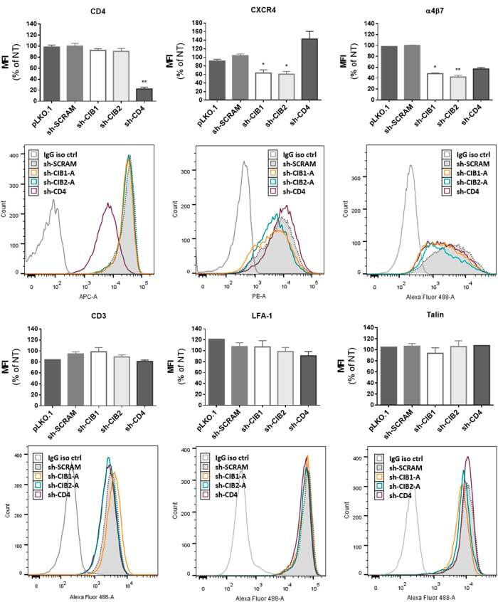
- Figure 9 Effect of downmodulation of CD4 and CIB proteins in CD4+ T-lymphocytes on the expression of proteins implicated in HIV-1 entry. Activated CD4+ T-lymphocytes were transduced with the empty vector (pLKO.1) or vectors leading to the expression of the indicated shRNAs, and the surface expression of the following molecules was measured by flow cytometry: CD3, CD4, CXCR4, LFA-1 and alpha4beta7. Intracellular talin expression was also evaluated in permeabilized cells. In each case, the mean fluorescence intensity (MFI) and a representative fluorescence histogram are shown. For MFI, the values represent the mean +- SEM of 4 independent experiments using two different shRNAs per targeted gene and are expressed relative to values obtained for non-transduced cells. *P < 0.05, **P < 0.01 versus sh-SCRAM (Kruskal-Wallis test).