Explore
 Validate
 Learn
Learn Immunohistochemistry
Antibody data
- Antibody Data
- Antigen structure
- References [5]
- Comments [0]
- Validations
- Immunohistochemistry [1]
- Other assay [4]
Submit
Validation data
Reference
Comment
Report error
- Product number
- 41-2444-80 - Provider product page

- Provider
- Invitrogen Antibodies
- Product name
- CD4 Monoclonal Antibody (N1UG0), eFluor™ 570, eBioscience™
- Antibody type
- Monoclonal
- Antigen
- Other
- Description
- Description: The N1UG0 monoclonal antibody reacts with human CD4, a 59 kDa cell surface glycoprotein expressed by the majority of thymocytes, a subpopulation of mature T cells (T-helper cells) and in low levels on monocytes. CD4 is a receptor for the human immunodeficiency virus (HIV). The N1UG0 antibody is recommended for use in staining human formalin-fixed paraffin embedded tissue sections. Applications Reported: This N1UG0 antibody has been reported for use in immunohistochemical staining of formalin-fixed paraffin embedded tissue sections. Applications Tested: This N1UG0 antibody has been tested by immunohistochemistry of formalin-fixed paraffin embedded human tissue using low or high pH antigen retrieval at less than or equal to 20 µg/mL. It is recommended that the antibody be titrated for opitmal performance in the assay of interest. Filter Recommendation: When using this eFluor® 570 antibody conjugate, we recommend a filter that will capture the 570 emission wavelength (for example, Excitation 545/25, 565LP, Emission 605/70). A standard Alexa Fluor® 555 or TRITC filter is acceptable. Excitation: 555 nm; Emission: 570 nm
- Reactivity
- Human
- Host
- Mouse
- Isotype
- IgG
- Antibody clone number
- N1UG0
- Vial size
- 25 µg
- Concentration
- 0.2 mg/mL
- Storage
- 4° C, store in dark, DO NOT FREEZE!
Submitted references Molecular Mechanism of Sphingosine-1-Phosphate Receptor 1 Regulating CD4(+) Tissue Memory in situ T Cells in Primary Sjogren's Syndrome.
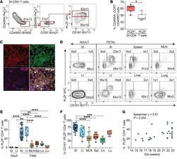
CD161 contributes to prenatal immune suppression of IFNγ-producing PLZF+ T cells.
Highly multiplexed immunofluorescence images and single-cell data of immune markers in tonsil and lung cancer.
Highly multiplexed immunofluorescence imaging of human tissues and tumors using t-CyCIF and conventional optical microscopes.
Inhibition of gp160 and CD4 maturation in U937 cells after both defective and productive infections by human immunodeficiency virus type 1.
Yang XX, Yang C, Wang L, Zhou YB, Yuan X, Xiang N, Wang YP, Li XM
International journal of general medicine 2021;14:6177-6188
International journal of general medicine 2021;14:6177-6188
CD161 contributes to prenatal immune suppression of IFNγ-producing PLZF+ T cells.
Halkias J, Rackaityte E, Hillman SL, Aran D, Mendoza VF, Marshall LR, MacKenzie TC, Burt TD
The Journal of clinical investigation 2019 May 30;129(9):3562-3577
The Journal of clinical investigation 2019 May 30;129(9):3562-3577
Highly multiplexed immunofluorescence images and single-cell data of immune markers in tonsil and lung cancer.
Rashid R, Gaglia G, Chen YA, Lin JR, Du Z, Maliga Z, Schapiro D, Yapp C, Muhlich J, Sokolov A, Sorger P, Santagata S
Scientific data 2019 Dec 17;6(1):323
Scientific data 2019 Dec 17;6(1):323
Highly multiplexed immunofluorescence imaging of human tissues and tumors using t-CyCIF and conventional optical microscopes.
Lin JR, Izar B, Wang S, Yapp C, Mei S, Shah PM, Santagata S, Sorger PK
eLife 2018 Jul 11;7
eLife 2018 Jul 11;7
Inhibition of gp160 and CD4 maturation in U937 cells after both defective and productive infections by human immunodeficiency virus type 1.
Bour S, Boulerice F, Wainberg MA
Journal of virology 1991 Dec;65(12):6387-96
Journal of virology 1991 Dec;65(12):6387-96
No comments: Submit comment
Supportive validation
- Submitted by
- Invitrogen Antibodies (provider)
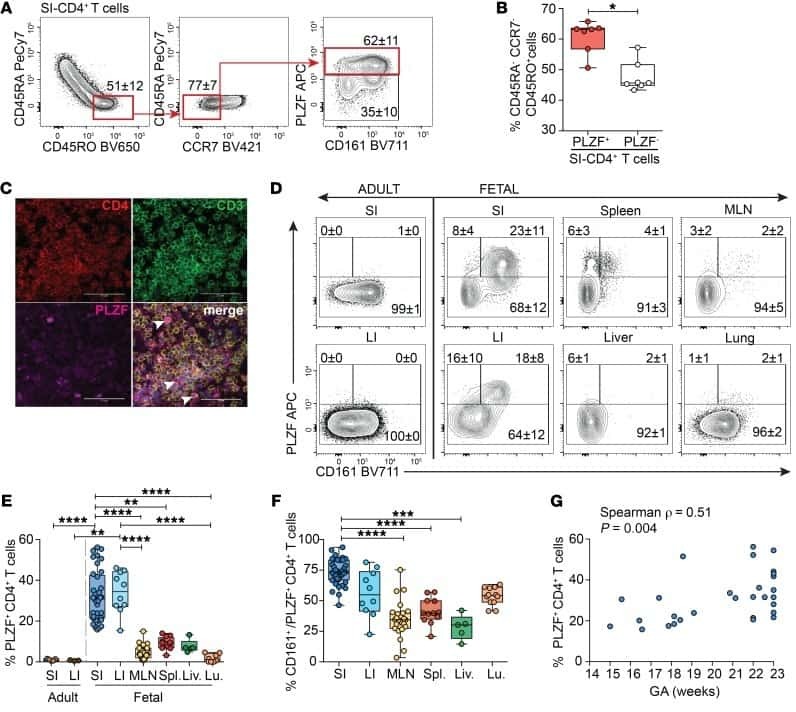
- Main image
- Experimental details
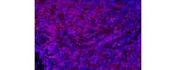
- Immunohistochemistry of formalin-fixed paraffin embedded human tonsil using 20 µg/mL of Anti-Human CD4 eFluor® 570. Nuclei are stained with DAPI.

Supportive validation
- Submitted by
- Invitrogen Antibodies (provider)
- Main image
- Experimental details
- NULL

- Submitted by
- Invitrogen Antibodies (provider)
- Main image

- Experimental details
- NULL
- Submitted by
- Invitrogen Antibodies (provider)
- Main image
- Experimental details
- NULL
- Submitted by
- Invitrogen Antibodies (provider)
- Main image
- Experimental details
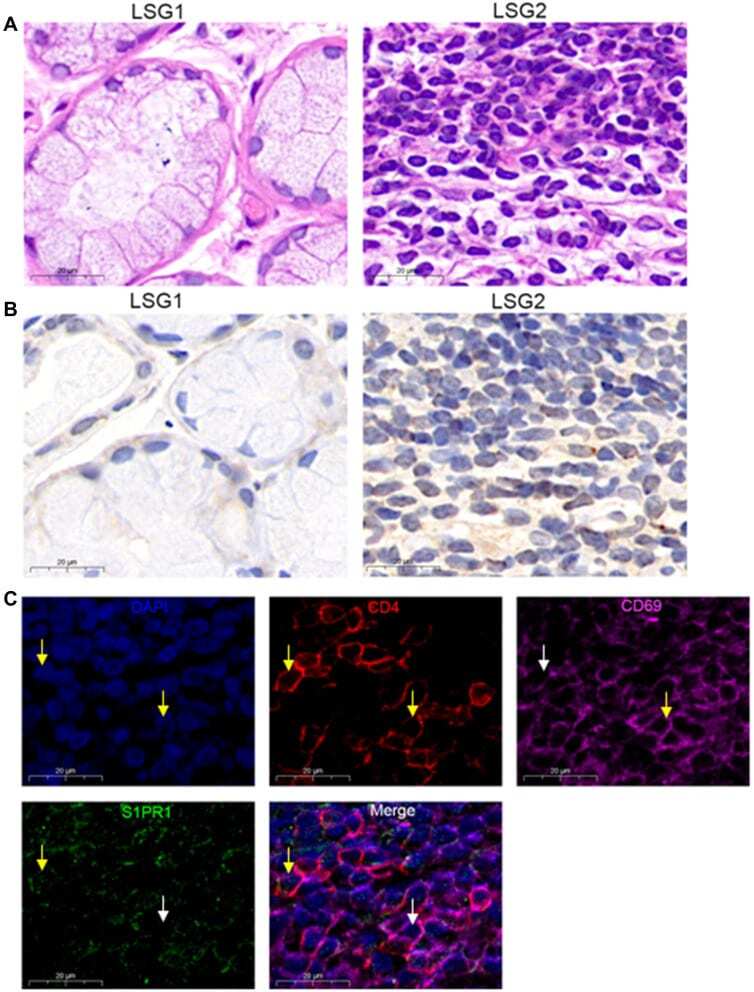
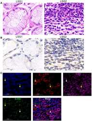
- Figure 1 S1PR1 expression in CD4 + CD69 + T RM cells. ( A ) Lymphocyte infiltration was detected by H&E staining of the labial salivary glands(LSG) of patients with pSS. ( B ) Expression of S1PR1 was detected by immunohistochemistry in labial salivary glands (LSG) of patients with pSS. ( C ) Immunofluorescence was performed to detect the expression of CD4, CD69 and S1PR1 in LSG samples with lymphocyte infiltration foci. Bar=20mum. DAPI was used to stain the nuclei and glowed blue. CD4 was pink light, CD69 was red light, and S1PR1 was green light. The yellow arrows point to positive fluorescence results and the white arrows point to negative fluorescence results in the DAPI, CD4, CD69 and S1PR1 image. In the in Merge image, the yellow arrows represent only S1PR1 expression in CD4 + T cells and no surface expression of CD69, the white arrows point to the CD4 + CD69 + T cells without the expression of S1PR1.