Explore
 Validate
 Learn
Learn Western blot
 Immunocytochemistry
 Flow cytometry
Antibody data
- Antibody Data
- Antigen structure
- References [1]
- Comments [0]
- Validations
- Immunocytochemistry [3]
- Immunohistochemistry [2]
- Other assay [4]
Submit
Validation data
Reference
Comment
Report error
- Product number
- MA5-32166 - Provider product page

- Provider
- Invitrogen Antibodies
- Product name
- CD4 Recombinant Rabbit Monoclonal Antibody (ST0488)
- Antibody type
- Monoclonal
- Antigen
- Recombinant full-length protein
- Description
- Recombinant rabbit monoclonal antibodies are produced using in vitro expression systems. The expression systems are developed by cloning in the specific antibody DNA sequences from immunoreactive rabbits. Then, individual clones are screened to select the best candidates for production. The advantages of using recombinant rabbit monoclonal antibodies include: better specificity and sensitivity, lot-to-lot consistency, animal origin-free formulations, and broader immunoreactivity to diverse targets due to larger rabbit immune repertoire.
- Reactivity
- Human
- Host
- Rabbit
- Isotype
- IgG
- Antibody clone number
- ST0488
- Vial size
- 100 μL
- Concentration
- 1 mg/mL
- Storage
- Store at 4°C short term. For long term storage, store at -20°C, avoiding freeze/thaw cycles.
Submitted references EMP3 mediates glioblastoma-associated macrophage infiltration to drive T cell exclusion.
Chen Q, Jin J, Huang X, Wu F, Huang H, Zhan R
Journal of experimental & clinical cancer research : CR 2021 May 8;40(1):160
Journal of experimental & clinical cancer research : CR 2021 May 8;40(1):160
No comments: Submit comment
Supportive validation
- Submitted by
- Invitrogen Antibodies (provider)
- Main image

- Experimental details
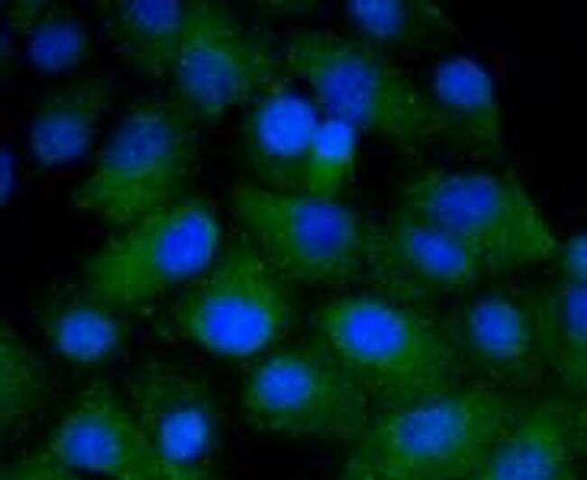
- Immunocytochemical analysis of CD4 in A549 cells using a CD4 Monoclonal antibody (Product # MA5-32166) as seen in green. The nuclear counter stain is DAPI (blue). Cells were fixed in paraformaldehyde, permeabilised with 0.25% Triton X100/PBS.
- Submitted by
- Invitrogen Antibodies (provider)
- Main image

- Experimental details
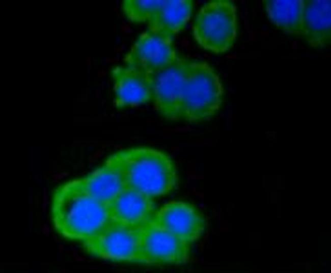
- Immunocytochemical analysis of CD4 in CRC cells using a CD4 Monoclonal antibody (Product # MA5-32166) as seen in green. The nuclear counter stain is DAPI (blue). Cells were fixed in paraformaldehyde, permeabilised with 0.25% Triton X100/PBS.

- Submitted by
- Invitrogen Antibodies (provider)
- Main image

- Experimental details
- Immunocytochemical analysis of CD4 in CRC cells using a CD4 Monoclonal antibody (Product # MA5-32166) as seen in green. The nuclear counter stain is DAPI (blue). Cells were fixed in paraformaldehyde, permeabilised with 0.25% Triton X100/PBS.
Supportive validation
- Submitted by
- Invitrogen Antibodies (provider)
- Main image
- Experimental details
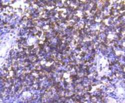
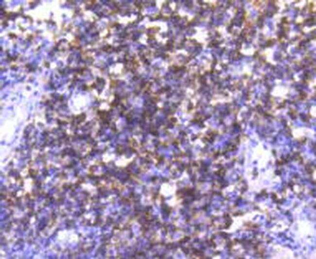
- Immunohistochemical analysis of CD4 of paraffin-embedded Human tonsil tissue using a CD4 Monoclonal antibody (Product #MA5-32166). Counter stained with hematoxylin.
- Submitted by
- Invitrogen Antibodies (provider)
- Main image
- Experimental details
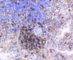
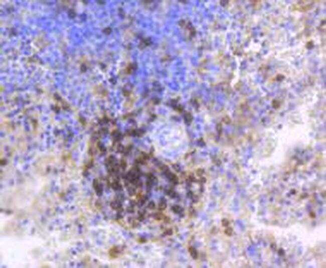
- Immunohistochemical analysis of CD4 of paraffin-embedded Human spleen tissue using a CD4 Monoclonal antibody (Product #MA5-32166). Counter stained with hematoxylin.
Supportive validation
- Submitted by
- Invitrogen Antibodies (provider)
- Main image
- Experimental details
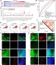
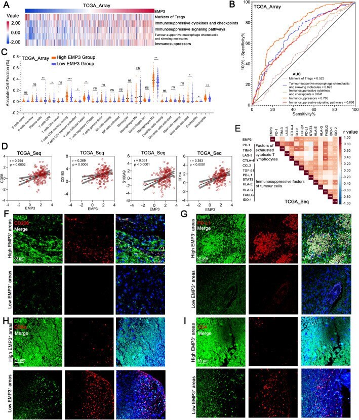
- Fig. 1 EMP3 is associated with immunosuppression in GBM. a Pearson correlation analysis of the expression of EMP3 with GBM-associated immunosuppressive metagenes in the TCGA_array GBM dataset ( n = 475). b ROC curves indicated that high EMP3 was involved in immunosuppression in the TCGA_array GBM dataset ( n = 475). c Immune cell fractions were estimated using CIBERSORT, and the differences between the cell fractions of the high and low EMP3 groups in the TCGA_array GBM dataset were evaluated using Student''s t-test ( n = 475). d Pearson correlation plots of the expression of EMP3 with M2 TAM signature markers (CD68, CD163, S100A9, and CD14) in the TCGA_array GBM dataset ( n = 475). e Correlation coefficient graph revealing the correlations of EMP3 with common immunosuppressive factors in the TCGA_seq GBM dataset ( n = 153). f Representative images of immunofluorescence (IF) staining for EMP3 and CD206 in different areas of serial sections from human GBM samples ( n = 27). Scale bar = 50 mum. g Representative images of IF staining for EMP3 and PD-L1 in different areas of serial sections of human GBM samples ( n = 27). Scale bar = 50 mum. h Representative images of IF staining for EMP3 and CD8alpha in different areas of serial sections from human GBM samples ( n = 27). Scale bar = 50 mum. I. Representative images of IF staining for EMP3 and CD4 in different areas of serial sections of human GBM samples ( n = 27). Scale bar = 50 mum. The mean +- S.D. is shown. Ns: nonsignifican
- Submitted by
- Invitrogen Antibodies (provider)
- Main image
- Experimental details
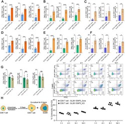
- Fig. 4 EMP3 drives T cell exclusion from the GBM microenvironment. a - c Percentages of IFN-gamma + /TNF-alpha + /IL-2 + CD4 + T cells in mouse tumours, blood, and spleens from the EMP3_KO and EMP3_Scra GL261 groups. Student''s t-test was performed. d - f Percentages of IFN-gamma + /TNF-alpha + /IL-2 + CD8 + T cells in mouse tumours, blood, and spleens from the EMP3_KO and EMP3_Scra GL261 groups. Data were analysed using Student''s t-test. g Percentages of FoxP3 + CD4 + T cells in mouse tumours, blood, and spleens from the EMP3_KO and EMP3_Scra GL261 groups. Student''s t-test was performed. h CD8 + T cells were isolated from splenocytes from C57BL/6 mice and activated with anti-CD3/CD28 Dynabeads for 3 days. Activated CD8 + T cells were co-cultured with EMP3_KO or EMP3_Scra GL261 cells at ratios of 1:1, 2:1, 5:1, and 10:1. Cell apoptosis analysis was conducted with the Annexin V-FITC Apoptosis Detection Kit (BD Biosciences, USA) according to the manufacturer''s instructions. Student''s t-test was performed. The mean +- S.D. is shown. Ns: nonsignificant, * p < 0.05, ** p < 0.01, and *** p < 0.001

- Submitted by
- Invitrogen Antibodies (provider)
- Main image
- Experimental details
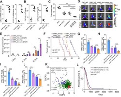
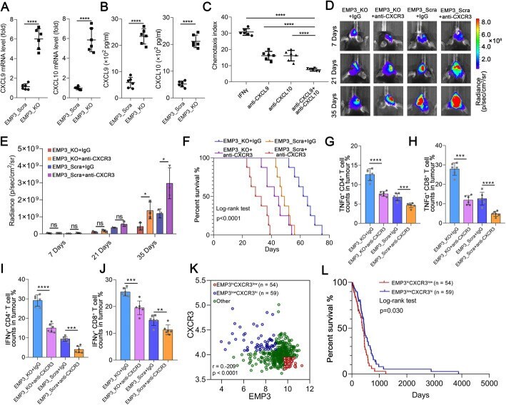
- Fig. 5 Loss of EMP3 in GBM cells promotes T cell infiltration via CXCL10 and CXCL9 in macrophages. a - b CXCL9 and CXCL10 mRNA and protein expression in RAW264.7 cells exposed to supernatants from EMP3_KO or EMP3_Scra GL261 cells for 48 hours was detected by qRT-PCR and ELISA. Student''s t-test was performed. KO: Knockout; Scra: Scramble. c Chemotaxis of T cells towards RAW264.7 cells treated with IFN-gamma, anti-CXCL9 and/or CXCL10. Student''s t-test was performed. d Bioluminescent images of C57BL/6 mice treated with an anti-alphaCXCR3 antibody in the EMP3_KO and EMP3_Scra groups on days 14, 21 and 35. KO: Knockout; Scra: Scramble. e Quantification of the bioluminescence imaging signal intensities in C57BL/6 mice. Student''s t-test was performed. f Kaplan-Meier survival curves of mice bearing intracranial EMP3_KO or EMP3_Scra GL261 tumours treated with the anti-CXCR3 antibody or isotype IgG. KO: Knockout; Scra: Scramble. The log-rank test was performed. g - h Percentages of TNFalpha + CD4 + and TNFalpha + CD8 + T cells in tumours from the EMP3_KO and EMP3_Scra GL261 groups treated with the anti-CXCR3 antibody or isotype IgG. Student''s t-test was performed. KO: Knockout; Scra: Scramble. i - j Percentages of IFN-gamma + CD4 + and IFN-gamma + CD8 + T cells in tumours from the EMP3_KO and EMP3_Scra GL261 groups treated with the anti-CXCR3 antibody or isotype IgG. Student''s t-test was performed. KO: Knockout; Scra: Scramble. k Pearson correlation plots of the expression of EMP3
- Submitted by
- Invitrogen Antibodies (provider)
- Main image

- Experimental details
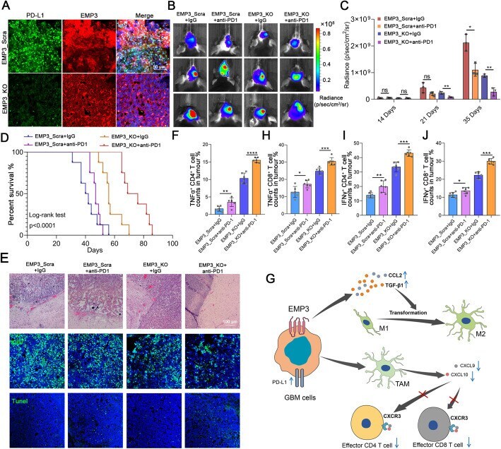
- Fig. 6 Knockout of EMP3 and immune checkpoint blockade have synergistic antitumorigenic effects. a Representative images of IF staining for EMP3 and PD-L1 in mouse brain tissues from the EMP3_KO and EMP3_Scra GL261 groups. Scale bar = 50 mum. KO: Knockout; Scra: Scramble. b Bioluminescence images of EMP3_KO and EMP3_Scra C57BL/6 mice treated with isotype IgG or PD1 blockade. KO: Knockout; Scra: Scramble. c Quantification of the bioluminescence imaging signal intensities in C57BL/6 mice. Student''s t-test was performed. d Kaplan-Meier survival curves of mice bearing intracranial EMP3_KO or EMP3_Scra GL261 tumours treated with an anti-PD1 antibody or isotype IgG. KO: Knockout; Scra: Scramble. The log-rank test was performed. e Tumour sections from the EMP3_KO and EMP3_Scra GL261 groups treated with the anti-PD1 antibody or isotype IgG were used for a TUNEL assay, IF staining and H&E analysis. Upper: Fluorescence images of TUNEL staining in tumours. Middle: Representative images of IF staining for Ki67 in tumours. Lower: H&E staining of tumour sections. Scale bar = 100 mum. f - h Percentages of TNFalpha + CD4 + and TNFalpha + CD8 + T cells in tumours from the EMP3_KO and EMP3_Scra GL261 groups treated with the anti-PD1 antibody or isotype IgG. Student''s t-test was performed. KO: Knockout; Scra: Scramble. i - j Percentages of IFN-gamma + CD4 + and IFN-gamma + CD8 + T cells in tumours from the EMP3_KO and EMP3_Scra GL261 groups treated with the anti-PD1 antibody or isotype IgG. S