Explore
 Validate
 Learn
Learn Western blot
Antibody data
- Antibody Data
- Antigen structure
- References [0]
- Comments [0]
- Validations
- Western blot [1]
- Immunohistochemistry [2]
- Flow cytometry [1]
Submit
Validation data
Reference
Comment
Report error
- Product number
- AP11517PU-N - Provider product page

- Provider
- Acris Antibodies GmbH
- Proper citation
- Acris Antibodies GmbH Cat#AP11517PU-N, RRID:AB_1750036
- Product name
- anti CD4 (C-term)
- Antibody type
- Polyclonal
- Antigen
- This antibody is generated from rabbits immunized with a KLH conjugated synthetic peptide between 336~366 from the C-terminal region of Human CD4.
- Reactivity
- Human
- Host
- Rabbit
- Vial size
- 0.4 ml
- Concentration
- lot specific
No comments: Submit comment
Supportive validation
- Submitted by
- Acris Antibodies GmbH (provider)
- Main image
- Experimental details
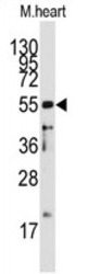
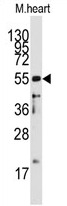
- Western blot analysis: AP11517PU-N CD4 Antibody staining of Mouse heart tissue lysates (35 µg/lane). CD4 (arrow) was detected using the purified Pab (1/60 dilution).
Supportive validation
- Submitted by
- Acris Antibodies GmbH (provider)
- Main image

- Experimental details
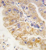
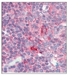
- Immunohistochemistry: AP11517PU-N CD4 antibody staining of Formalin-Fixed, Paraffin-Embedded Human lung carcinoma tissue. This antibody was peroxidase-conjugated to the secondary antibody, followed by DAB staining. This data demonstrates the use of this antibody for immunohistochemistry. Clinical relevance has not been evaluated.
- Submitted by
- Acris Antibodies GmbH (provider)
- Main image
- Experimental details
- Immunohistochemistry: AP11517PU-N CD4 antibody staining of Formalin-Fixed, Paraffin-Embedded Human Tonsil tissue. This antibody was peroxidase-conjugated to the secondary antibody, followed by DAB staining. This data demonstrates the use of this antibody for immunohistochemistry. Clinical relevance has not been evaluated.

Supportive validation
- Submitted by
- Acris Antibodies GmbH (provider)
- Main image
- Experimental details
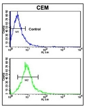
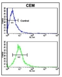
- Flow cytometric analysis of CEM cells using AP11517PU-N CD4 Antibody (C-term) (bottom histogram) compared to a Negative Control cell (top histogram). FITC-conjugated goat-anti-rabbit secondary antibodies were used for the analysis.