 Explore
 Validate
 Learn
Learn Western blot
Antibody data
- Antibody Data
- Antigen structure
- References [1]
- Comments [0]
- Validations
- Western blot [1]
- ELISA [1]
- Immunohistochemistry [1]
Submit
Validation data
Reference
Comment
Report error
- Product number
- AF6240 - Provider product page

- Provider
- R&D Systems
- Product name
- Human Prostatic Acid Phosphatase/ACPP Antibody
- Antibody type
- Polyclonal
- Description
- Antigen Affinity-purified. Detects human Prostatic Acid Phosphatase/ACPP in direct ELISAs and Western blots.
- Reactivity
- Human
- Host
- Sheep
- Conjugate
- Unconjugated
- Antigen sequence
P15309- Isotype
- IgG
- Vial size
- 100 ug
- Concentration
- LYOPH
- Storage
- Use a manual defrost freezer and avoid repeated freeze-thaw cycles. 12 months from date of receipt, -20 to -70 °C as supplied. 1 month, 2 to 8 °C under sterile conditions after reconstitution. 6 months, -20 to -70 °C under sterile conditions after reconstitution.
Submitted references Up-regulated Ectonucleotidases in Fas-Associated Death Domain Protein- and Receptor-Interacting Protein Kinase 1-Deficient Jurkat Leukemia Cells Counteract Extracellular ATP/AMP Accumulation via Pannexin-1 Channels during Chemotherapeutic Drug-Induced Apoptosis.
Boyd-Tressler AM, Lane GS, Dubyak GR
Molecular pharmacology 2017 Jul;92(1):30-47
Molecular pharmacology 2017 Jul;92(1):30-47
No comments: Submit comment
Supportive validation
- Submitted by
- R&D Systems (provider)
- Main image
- Experimental details
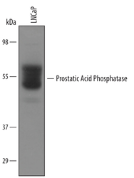
- Detection of Human Prostatic Acid Phosphatase/ACPP by Western Blot. Western blot shows lysates of LNCaP human prostate cancer cell line. PVDF Membrane was probed with 1 µg/mL of Human Prostatic Acid Phosphatase/ACPP Antigen Affinity-purified Polyclonal Antibody (Catalog # AF6240) followed by HRP-conjugated Anti-Sheep IgG Secondary Antibody (Catalog # HAF016). A specific band was detected for Prostatic Acid Phosphatase/ACPP at approximately 50-55 kDa (as indicated). This experiment was conducted under reducing conditions and using Immunoblot Buffer Group 8.

Supportive validation
- Submitted by
- R&D Systems (provider)
- Main image
- Experimental details
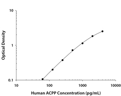
- Human Prostatic Acid Phosphatase/ACPP ELISA Standard Curve. Recombinant Human Prostatic Acid Phosphatase/ACPP protein was serially diluted 2-fold and captured by Mouse Anti-Human Prostatic Acid Phosphatase/ACPP Monoclonal Antibody (Catalog # MAB62401) coated on a Clear Polystyrene Microplate (Catalog # DY990). Sheep Anti-Human Prostatic Acid Phosphatase/ACPP Antigen Affinity-purified Polyclonal Antibody (Catalog # AF6240) was biotinylated and incubated with the protein captured on the plate. Detection of the standard curve was achieved by incubating Streptavidin-HRP (Catalog # DY998) followed by Substrate Solution (Catalog # DY999) and stopping the enzymatic reaction with Stop Solution (Catalog # DY994).
Supportive validation
- Submitted by
- R&D Systems (provider)
- Main image
- Experimental details
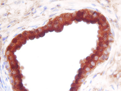
- Prostatic Acid Phosphatase/ACPP in Human Prostate. Prostatic Acid Phosphatase/ACPP was detected in immersion fixed paraffin-embedded sections of human prostate using Human Prostatic Acid Phosphatase/ACPP Antigen Affinity-purified Polyclonal Antibody (Catalog # AF6240) at 3 µg/mL overnight at 4 °C. Before incubation with the primary antibody, tissue was subjected to heat-induced epitope retrieval using Antigen Retrieval Reagent-Basic (Catalog # CTS013). Tissue was stained using the Anti-Sheep HRP-DAB Cell & Tissue Staining Kit (brown; Catalog # CTS019) and counterstained with hemotoxylin (blue). Specific staining was localized to the cytoplasm of epithelial cells. View our protocol for Chromogenic IHC Staining of Paraffin-embedded Tissue Sections.
