Explore
 Validate
 Learn
Learn Western blot
 Immunocytochemistry
Antibody data
- Antibody Data
- Antigen structure
- References [1]
- Comments [0]
- Validations
- Immunocytochemistry [2]
- Immunohistochemistry [1]
- Other assay [1]
Submit
Validation data
Reference
Comment
Report error
- Product number
- PA5-52115 - Provider product page

- Provider
- Invitrogen Antibodies
- Product name
- Vezatin Polyclonal Antibody
- Antibody type
- Polyclonal
- Antigen
- Recombinant protein fragment
- Description
- Immunogen sequence: DELEKLVCTK ETQELVSEAY PILEQKLKLI QPHVQASNNC WEEAISQVDK LLRRNTDKKG KPEIACENPH CTVVPLKQPT LHIADKDPIP EEQELEAYVD DIDIDSDFRK DDFYYLSQED KERQKREHEE SKRVLQELKS VLG Highest antigen sequence identity to the following orthologs: Mouse - 87%, Rat - 83%.
- Reactivity
- Human
- Host
- Rabbit
- Isotype
- IgG
- Vial size
- 100 μL
- Concentration
- 0.3 mg/mL
- Storage
- Store at 4°C short term. For long term storage, store at -20°C, avoiding freeze/thaw cycles.
Submitted references In situ molecular characterization of endoneurial microvessels that form the blood-nerve barrier in normal human adult peripheral nerves.
Ouyang X, Dong C, Ubogu EE
Journal of the peripheral nervous system : JPNS 2019 Jun;24(2):195-206
Journal of the peripheral nervous system : JPNS 2019 Jun;24(2):195-206
No comments: Submit comment
Supportive validation
- Submitted by
- Invitrogen Antibodies (provider)
- Main image

- Experimental details
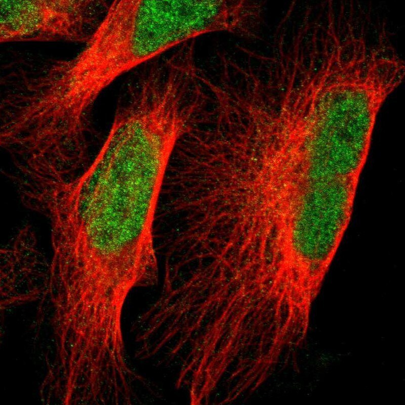
- Immunofluorescent staining of Vezatin in human cell line U-2 OS shows positivity in nucleus but excluded from the nucleoli. Samples were probed using a Vezatin Polyclonal Antibody (Product # PA5-52115).

- Submitted by
- Invitrogen Antibodies (provider)
- Main image

- Experimental details
- Immunofluorecent analysis of Vezatin in human cell line U-2 OS using Vezatin Polyclonal Antibody (Product # PA5-52115). Staining shows localization to nucleoplasm.
Supportive validation
- Submitted by
- Invitrogen Antibodies (provider)
- Main image
- Experimental details
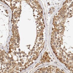
- Immunohistochemical staining of Vezatin in human testis using a Vezatin Polyclonal Antibody (Product # PA5-52115) shows moderate cytoplasmic positivity in cells in seminiferous ducts and Leydig cells.

Supportive validation
- Submitted by
- Invitrogen Antibodies (provider)
- Main image
- Experimental details
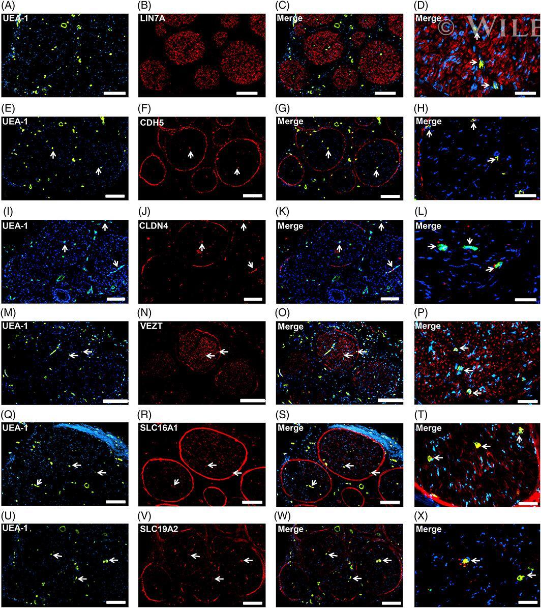
- Restrictive junctional complex and transporter proteins. Representative indirect fluorescent digital photomicrographs of cryostat axial sections of normal adult sural nerves show UEA-1-positive endothelial cells (green; A, E, I, M, Q, U) with expression of LIN7A, CDH5, CLDN4, VEZT, SLC16A1 and SLC19A2 (red; B, F, J, N, R, V, respectively) restricted to endoneurial microvessels shown in the merged images at lower and higher magnifications (yellow/ orange). LIN7A expression by endoneurial microvessels is apparent only at higher magnification (D). These proteins are expressed by the restrictive blood-nerve barrier and shared with other selected cell types suggesting specialized roles in the restrictive junctional complex formation (LIN7A, CDH5, CLDN4 and VEZT) or specialized nutrient transporters (SLC16A1 and SLC19A2) within normal adult peripheral nerves, as determined by known morphological profiles in situ. White arrows demonstrate positively staining endoneurial microvascular endothelium. Blue (4, 6-diamidino-2-phenylindole) staining indicates nuclei. Scale bar 500 mum for A-C, E-G, I-K, M-O, Q-S and U-W, and 125 mum for D, H, L, P, T and X