Explore
 Validate
 Learn
Learn Western blot
 Immunocytochemistry
Antibody data
- Antibody Data
- Antigen structure
- References [3]
- Comments [0]
- Validations
- Immunocytochemistry [8]
- Immunohistochemistry [4]
- Other assay [2]
Submit
Validation data
Reference
Comment
Report error
- Product number
- PA5-28195 - Provider product page

- Provider
- Invitrogen Antibodies
- Product name
- OGDH Polyclonal Antibody
- Antibody type
- Polyclonal
- Antigen
- Recombinant full-length protein
- Description
- Recommended positive controls: 293T, A431, HeLa, HepG2, Huh-7, Mouse brain. Predicted reactivity: Mouse (93%), Rat (93%), Xenopus laevis (84%), Chicken (87%), Bovine (97%). Store product as a concentrated solution. Centrifuge briefly prior to opening the vial.
- Reactivity
- Human, Mouse
- Host
- Rabbit
- Isotype
- IgG
- Vial size
- 100 μL
- Concentration
- 0.3 mg/mL
- Storage
- Store at 4°C short term. For long term storage, store at -20°C, avoiding freeze/thaw cycles.
Submitted references Quantitative Proteomic Approach Reveals Altered Metabolic Pathways in Response to the Inhibition of Lysine Deacetylases in A549 Cells under Normoxia and Hypoxia.
Selective knockdown of hexokinase 2 in rods leads to age-related photoreceptor degeneration and retinal metabolic remodeling.
The Eating-Disorder Associated HDAC4(A778T) Mutation Alters Feeding Behaviors in Female Mice.
Martín-Bernabé A, Tarragó-Celada J, Cunin V, Michelland S, Cortés R, Poignant J, Boyault C, Rachidi W, Bourgoin-Voillard S, Cascante M, Seve M
International journal of molecular sciences 2021 Mar 25;22(7)
International journal of molecular sciences 2021 Mar 25;22(7)
Selective knockdown of hexokinase 2 in rods leads to age-related photoreceptor degeneration and retinal metabolic remodeling.
Zhang R, Shen W, Du J, Gillies MC
Cell death & disease 2020 Oct 20;11(10):885
Cell death & disease 2020 Oct 20;11(10):885
The Eating-Disorder Associated HDAC4(A778T) Mutation Alters Feeding Behaviors in Female Mice.
Lutter M, Khan MZ, Satio K, Davis KC, Kidder IJ, McDaniel L, Darbro BW, Pieper AA, Cui H
Biological psychiatry 2017 May 1;81(9):770-777
Biological psychiatry 2017 May 1;81(9):770-777
No comments: Submit comment
Supportive validation
- Submitted by
- Invitrogen Antibodies (provider)
- Main image
- Experimental details
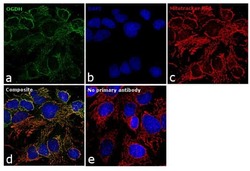
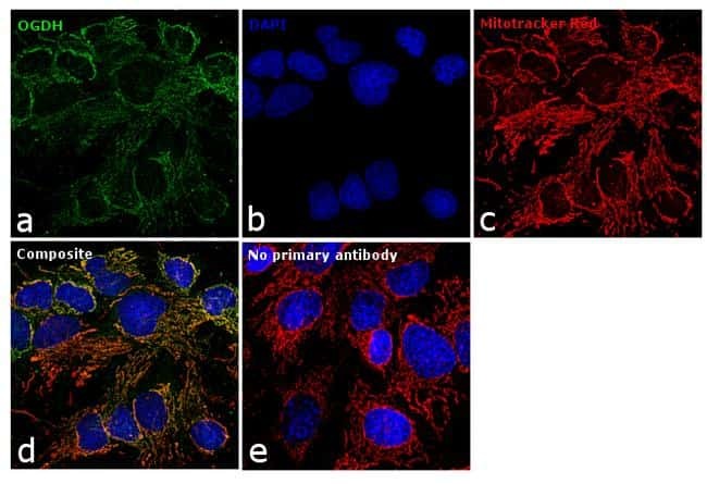
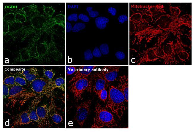
- Immunofluorescence analysis of OGDH was performed using 70% confluent log phase Hep G2 cells. The cells were fixed with 4% paraformaldehyde for 10 minutes, permeabilized with 0.1% Triton™ X-100 for 15 minutes, and blocked with 1% BSA for 1 hour at room temperature. The cells were labeled with OGDH Polyclonal Antibody (Product # PA5-28195) at 1:100 dilution in 0.1% BSA, incubated at 4 degree Celsius overnight and then labeled with Goat anti-Rabbit IgG (H+L) Superclonal™ Secondary Antibody, Alexa Fluor® 488 conjugate (Product # A27034) at a dilution of 1:2000 for 45 minutes at room temperature (Panel a: green). Nuclei (Panel b: blue) were stained with SlowFade® Gold Antifade Mountant with DAPI (Product # S36938). Mitochondria (Panel c: red) was stained with Mitotracker Red CMXRos (Product # M7512). Panel d represents the merged image showing mitochondrial localization. Panel e represents control cells with no primary antibody to assess background. The images were captured at 60X magnification.
- Submitted by
- Invitrogen Antibodies (provider)
- Main image

- Experimental details
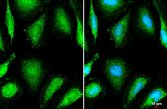
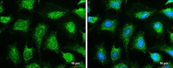
- Immunocytochemistry-Immunofluorescence analysis of OGDH was performed in HeLa cells fixed in ice-cold MeOH for 5 min. Green: OGDH Polyclonal Antibody (Product # PA5-28195) diluted at 1:500. Blue: Hoechst 33342 staining. Scale bar = 10 µm.

- Submitted by
- Invitrogen Antibodies (provider)
- Main image

- Experimental details
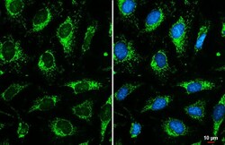
- OGDH Polyclonal Antibody detects OGDH protein at mitochondria by immunofluorescent analysis. Sample: HeLa cells were fixed in ice-cold MeOH for 5 min. Green: OGDH stained by OGDH Polyclonal Antibody (Product # PA5-28195) diluted at 1:200. Blue: Fluoroshield with DAPI . Scale bar= 10 µm.

- Submitted by
- Invitrogen Antibodies (provider)
- Main image

- Experimental details
- OGDH Polyclonal Antibody detects OGDH protein at mitochondria by immunofluorescent analysis. Sample: HeLa cells were fixed in ice-cold MeOH for 5 min. Green: OGDH stained by OGDH Polyclonal Antibody (Product # PA5-28195) diluted at 1:200. Blue: Fluoroshield with DAPI . Scale bar= 10 µm.
- Submitted by
- Invitrogen Antibodies (provider)
- Main image
- Experimental details
- Immunocytochemistry-Immunofluorescence analysis of OGDH was performed in HeLa cells fixed in ice-cold MeOH for 5 min. Green: OGDH Polyclonal Antibody (Product # PA5-28195) diluted at 1:500. Blue: Hoechst 33342 staining. Scale bar = 10 µm.

- Submitted by
- Invitrogen Antibodies (provider)
- Main image
- Experimental details
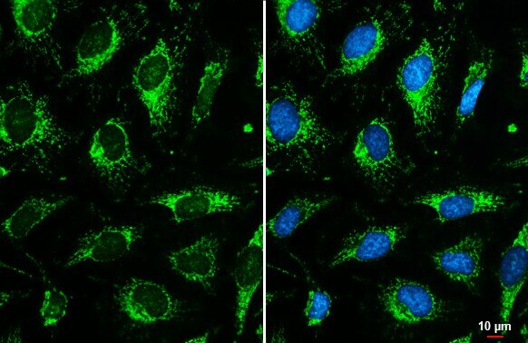
- Immunocytochemistry-Immunofluorescence analysis of OGDH was performed in HeLa cells fixed in ice-cold MeOH for 5 min. Green: OGDH Polyclonal Antibody (Product # PA5-28195) diluted at 1:500. Blue: Hoechst 33342 staining. Scale bar = 10 µm.
- Submitted by
- Invitrogen Antibodies (provider)
- Main image

- Experimental details
- Immunofluorescence analysis of OGDH was performed using 70% confluent log phase Hep G2 cells. The cells were fixed with 4% paraformaldehyde for 10 minutes, permeabilized with 0.1% Triton™ X-100 for 15 minutes, and blocked with 1% BSA for 1 hour at room temperature. The cells were labeled with OGDH Polyclonal Antibody (Product # PA5-28195) at 1:100 dilution in 0.1% BSA, incubated at 4 degree Celsius overnight and then labeled with Goat anti-Rabbit IgG (Heavy Chain) Superclonal™ Secondary Antibody, Alexa Fluor® 488 conjugate (Product # A27034) at a dilution of 1:2000 for 45 minutes at room temperature (Panel a: green). Nuclei (Panel b: blue) were stained with SlowFade® Gold Antifade Mountant with DAPI (Product # S36938). Mitochondria (Panel c: red) was stained with Mitotracker Red CMXRos (Product # M7512). Panel d represents the merged image showing mitochondrial localization. Panel e represents control cells with no primary antibody to assess background. The images were captured at 60X magnification.
Supportive validation
- Submitted by
- Invitrogen Antibodies (provider)
- Main image
- Experimental details
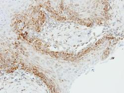
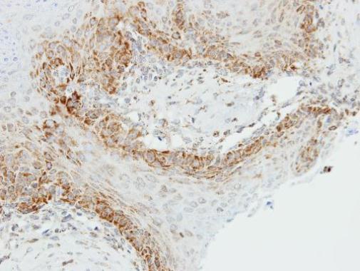
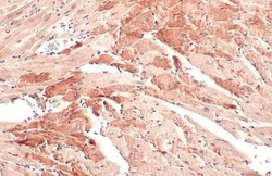
- Immunohistochemical analysis of paraffin-embedded Cal27 xenograft, using OGDH (Product # PA5-28195) antibody at 1:500 dilution. Antigen Retrieval: EDTA based buffer, pH 8.0, 15 min.
- Submitted by
- Invitrogen Antibodies (provider)
- Main image
- Experimental details
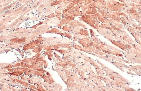
- OGDH Polyclonal Antibody detects OGDH protein at cytoplasm by immunohistochemical analysis. Sample: Paraffin-embedded mouse muscle. OGDH stained by OGDH Polyclonal Antibody (Product # PA5-28195) diluted at 1:500. Antigen Retrieval: Citrate buffer, pH 6.0, 15 min.
- Submitted by
- Invitrogen Antibodies (provider)
- Main image
- Experimental details
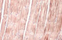
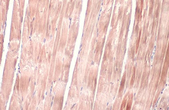
- OGDH Polyclonal Antibody detects OGDH protein at cytoplasm by immunohistochemical analysis. Sample: Paraffin-embedded mouse heart. OGDH stained by OGDH Polyclonal Antibody (Product # PA5-28195) diluted at 1:500. Antigen Retrieval: Citrate buffer, pH 6.0, 15 min.
- Submitted by
- Invitrogen Antibodies (provider)
- Main image
- Experimental details
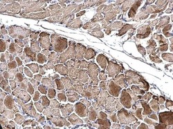
- OGDH Polyclonal Antibody detects OGDH protein at mitochondria on mouse muscle by immunohistochemical analysis. Sample: Paraffin-embedded mouse muscle. OGDH Polyclonal Antibody (Product # PA5-28195) dilution: 1:500. Antigen Retrieval: EDTA based buffer, pH 8.0, 15 min.

Supportive validation
- Submitted by
- Invitrogen Antibodies (provider)
- Main image
- Experimental details
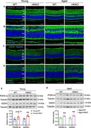
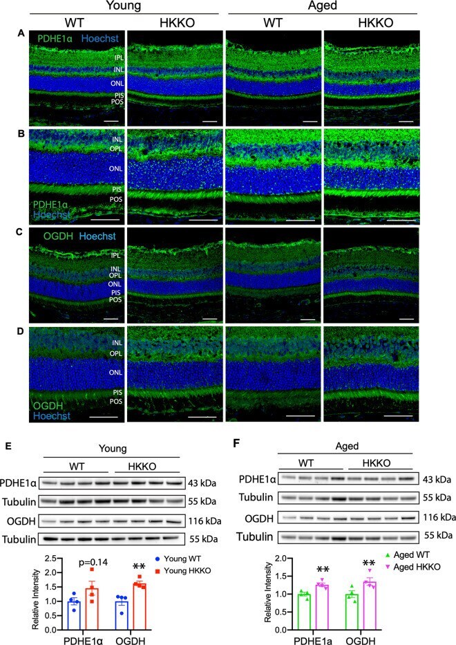
- Fig. 6 Knocking down HK2 led to upregulation of enzymes in the TCA cycle. A - D Immunostaining for enzymes in the TCA cycle including pyruvate dehydrogenase E1alpha (PDHE1alpha, A and B ) and oxoglutarate dehydrogenase (OGDH, C and D ) in retinal sections from HKKO mice and age-matched WT controls. Loss of HK2 in rods led to increased expression of PDHE1alpha ( A , B ) and OGDH ( C , D ) in the photoreceptor inner segments (PIS). The panel of images in ( B ) and ( D ) are higher-power images of areas in ( A ) and ( C ) respectively. Scale bar: 50 mum in ( A - D ). E , F Western blots indicated that the expression of PDHE1alpha ( C ) and OGDH ( D ) was significantly increased in HKKO mice compared with age-matched WT controls. * p < 0.05 and ** p < 0.01, n = 4/group.
- Submitted by
- Invitrogen Antibodies (provider)
- Main image
- Experimental details
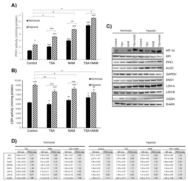
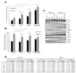
- Figure 4 Effect of KDAC inhibition on enzyme activities in A549 cells under normoxia and hypoxia. ( A , B ) The ATP- dependent 6-phosphofructokinase (PFK1) ( A ) and lactate dehydrogenase (LDH) ( B ) enzymatic activities were measured after 24 h of incubation, and activities were normalized to intracellular protein content in each condition. A549 cells were treated with 1 muM of TSA, 20 mM of NAM, and both 1 muM TSA and 20 mM NAM for 24 h of incubation under normoxia and hypoxia. Cells incubated in medium without KDACIs served as control. Bars represent the means +- standard error of the mean of three independent experiments. The asterisks above bars indicate statistically significant differences compared to normoxic control cells. The asterisks above curly brackets indicate statistically significant differences between hypoxic and normoxic treatments and between hypoxic treatments and hypoxic control cells. Statistical significance was assessed by a two-tailed Student''s t -test. *, p