Explore
 Validate
 Learn
Learn Western blot
Antibody data
- Antibody Data
- Antigen structure
- References [2]
- Comments [0]
- Validations
- Western blot [1]
- Immunocytochemistry [2]
- Other assay [2]
Submit
Validation data
Reference
Comment
Report error
- Product number
- MA5-27638 - Provider product page

- Provider
- Invitrogen Antibodies
- Product name
- Versican Monoclonal Antibody (N351/23)
- Antibody type
- Monoclonal
- Antigen
- Other
- Description
- 1 µg/mL of MA5-27638 was sufficient for detection of Versican in 20 µg of mouse brain membrane lysate and assayed by colorimetric immunoblot analysis using goat anti-mouse IgG:HRP as the secondary antibody.|Detects >350kDa. This antibody was formerly sold as clone S351-23
- Reactivity
- Human, Mouse, Rat
- Host
- Mouse
- Isotype
- IgG
- Antibody clone number
- N351/23
- Vial size
- 100 μg
- Concentration
- 1 mg/mL
- Storage
- -20°C
Submitted references The antifibrotic adipose-derived stromal cell: Grafted fat enriched with CD74+ adipose-derived stromal cells reduces chronic radiation-induced skin fibrosis.
Culture and Differentiation of Human Hair Follicle Dermal Papilla Cells in a Soft 3D Self-Assembling Peptide Scaffold.
Borrelli MR, Patel RA, Adem S, Diaz Deleon NM, Shen AH, Sokol J, Yen S, Chang EY, Nazerali R, Nguyen D, Momeni A, Wang KC, Longaker MT, Wan DC
Stem cells translational medicine 2020 Nov;9(11):1401-1413
Stem cells translational medicine 2020 Nov;9(11):1401-1413
Culture and Differentiation of Human Hair Follicle Dermal Papilla Cells in a Soft 3D Self-Assembling Peptide Scaffold.
Betriu N, Jarrosson-Moral C, Semino CE
Biomolecules 2020 Apr 28;10(5)
Biomolecules 2020 Apr 28;10(5)
No comments: Submit comment
Supportive validation
- Submitted by
- Invitrogen Antibodies (provider)
- Main image
- Experimental details
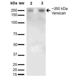
- Western blot analysis of Versican in rat brain with 15 µg of sample. The sample was blocked with 5% skim milk in TBST, incubated with Versican monoclonal antibody (Product # MA5-27638) using a dilution of 1:200 (16 hours at RT), followed by Goat Anti-Mouse (1 hour at RT) at a dilution of 1:1000 and TMB development (10 min at RT). Samples were arranged as follows: Lane 1: Molecular Weight Ladder, Lane 2: Rat Brain membrane and brain.

Supportive validation
- Submitted by
- Invitrogen Antibodies (provider)
- Main image

- Experimental details
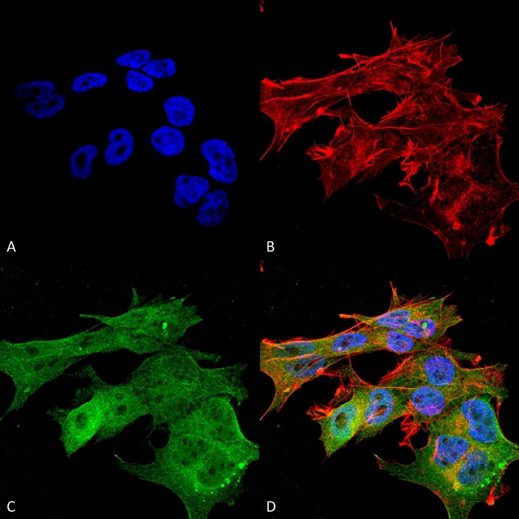
- Immunofluorescent analysis of Versican in human neuroblastoma cell line (SK-N-BE). Sample was fixed with 4% formaldehyde (15 min at RT), incubated with Versican monoclonal antibody (Product # MA5-27638) using a dilution of 1:100 (1 hour at RT), and followed by Goat Anti-Mouse 488, Phalloidin Texas Red and DAPI secondary antibody at a dilution of 1:100, 1:1000 (60 min at RT) and 1:5000 (5 min at RT). Images are shown as follows: (A) DAPI (blue) nuclear stain, B) Phalloidin Texas Red F-Actin stain, C) Versican Antibody, D) Merged image. Magnification: 60x.

- Submitted by
- Invitrogen Antibodies (provider)
- Main image

- Experimental details
- Immunofluorescent analysis of Versican in human neuroblastoma cell line (SK-N-BE). Sample was fixed with 4% formaldehyde (15 min at RT), incubated with Versican monoclonal antibody (Product # MA5-27638) using a dilution of 1:100 (1 hour at RT), and followed by Goat Anti-Mouse 488, Phalloidin Texas Red and DAPI secondary antibody at a dilution of 1:100, 1:1000 (60 min at RT) and 1:5000 (5 min at RT). Images are shown as follows: (A) DAPI (blue) nuclear stain, B) Phalloidin Texas Red F-Actin stain, C) Versican Antibody, D) Merged image. Magnification: 60x.
Supportive validation
- Submitted by
- Invitrogen Antibodies (provider)
- Main image
- Experimental details
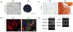
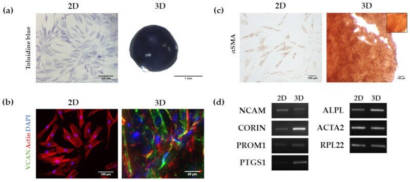
- Figure 6 Analysis of HFDPC markers in 2D and RAD16-I 3D cultures. ( a ) Toluidine blue staining for the detection of glycosaminoglycans; ( b ) Versican (VCAN) immunofluorescence. VCAN was absent in 2D cultures but its expression was recovered in 3D cultures as shown by immunofluorescence detection; ( c ) alpha Smooth Muscle Actin (alphaSMA) immunocytochemistry. alphaSMA was detected in both 2D and 3D cultures. The inner box represents a negative control (incubation with secondary antibody only); ( d ) Agarose gel of Reverse-transcription PCR products including Neural Cell Adhesion Molecule ( NCAM ), Corin ( CORIN ), Prominin-1 ( PROM1 ), Prostaglandin endoperoxidase synthase 1 ( PTGS1 ), alkaline phosphatase ( ALPL ) and alpha-Smooth Muscle Actin ( ACTA2 ).
- Submitted by
- Invitrogen Antibodies (provider)
- Main image
- Experimental details
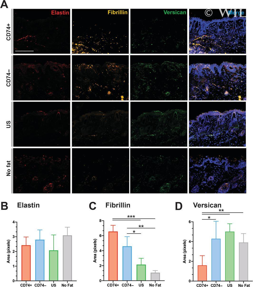
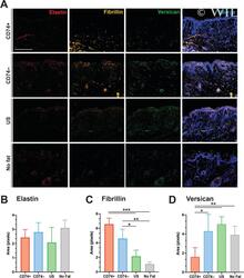
- 5 FIGURE Immunofluorescence staining for elastic fibers. A, Staining for elastin (red, far left column), fibrillin (yellow, second column), and versican (green, third column) highlights how fat grafting alters components of irradiated extracellular matrix. Merged image with 4',6-diamidino-2-phenylindole (DAPI) counterstain (blue) on the far-right column. B, Pixel positive-percent quantification of elastin staining, C, fibrillin staining, and, D, versican staining. Note mice grafted with fat enriched with CD74+ ASCs had improvement in staining for fibrillin (*** P < .001, ** P < .01, * P < .05), with a concomitant decrease of versican (** P < .01, * P < .05). Scale bar = 100 mum. ASC, adipose-derived stromal cell