Explore
 Validate
 Learn
Learn Western blot
 Immunohistochemistry
Antibody data
- Antibody Data
- Antigen structure
- References [1]
- Comments [0]
- Validations
- Immunohistochemistry [4]
- Other assay [3]
Submit
Validation data
Reference
Comment
Report error
- Product number
- PA5-54637 - Provider product page

- Provider
- Invitrogen Antibodies
- Product name
- LLGL1 Polyclonal Antibody
- Antibody type
- Polyclonal
- Antigen
- Recombinant protein fragment
- Description
- Immunogen sequence: FERFSLSARN ITEPLCSLDI NWPRDATQAS YRIRESPKLS QANGTPSILL APQSLDGSPD PAHSMGPDTP EPPEAALSPM SIDSATSADT TLDTTGDVTV EDVKDFLGSS EESEKNLRNL AEDE Highest antigen sequence identity to the following orthologs: Mouse - 76%, Rat - 72%.
- Reactivity
- Human
- Host
- Rabbit
- Isotype
- IgG
- Vial size
- 100 μL
- Concentration
- 0.4 mg/mL
- Storage
- Store at 4°C short term. For long term storage, store at -20°C, avoiding freeze/thaw cycles.
Submitted references Scribble, Erbin, and Lano redundantly regulate epithelial polarity and apical adhesion complex.
Choi J, Troyanovsky RB, Indra I, Mitchell BJ, Troyanovsky SM
The Journal of cell biology 2019 Jul 1;218(7):2277-2293
The Journal of cell biology 2019 Jul 1;218(7):2277-2293
No comments: Submit comment
Supportive validation
- Submitted by
- Invitrogen Antibodies (provider)
- Main image
- Experimental details
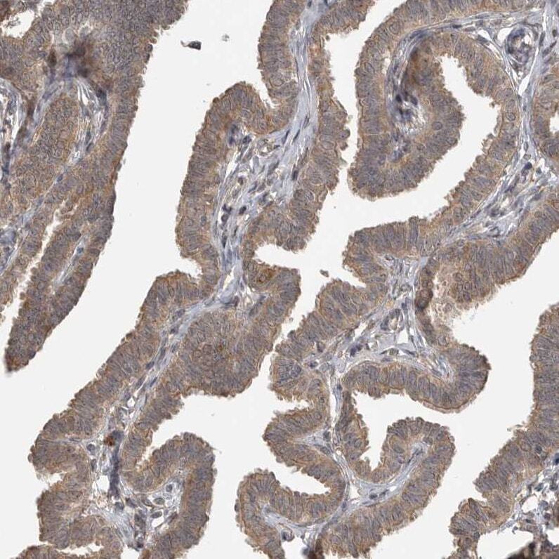
- Immunohistochemical analysis of LLGL1 in human stomach using LLGL1 Polyclonal Antibody (Product # PA5-54637) shows strong cytoplasmic and membranous positivity in glandular cells.

- Submitted by
- Invitrogen Antibodies (provider)
- Main image

- Experimental details
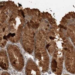
- Immunohistochemical analysis of LLGL1 in human fallopian tube using LLGL1 Polyclonal Antibody (Product # PA5-54637) shows moderate cytoplasmic positivity in glandular cells.
- Submitted by
- Invitrogen Antibodies (provider)
- Main image
- Experimental details
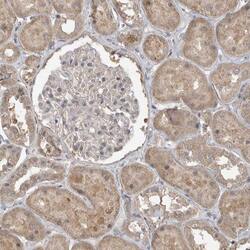
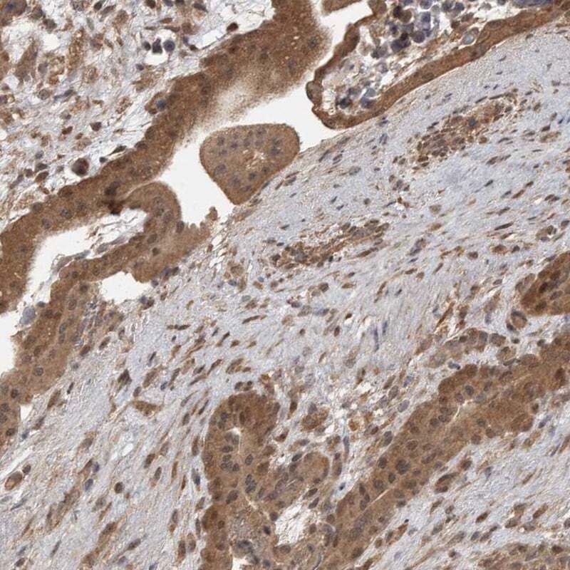
- Immunohistochemical analysis of LLGL1 in human kidney using LLGL1 Polyclonal Antibody (Product # PA5-54637) shows weak cytoplasmic positivity in cells in tubules.

- Submitted by
- Invitrogen Antibodies (provider)
- Main image

- Experimental details
- Immunohistochemical staining of LLGL1 in human placenta using a LLGL1 Polyclonal Antibody (Product # PA5-54637) shows moderate cytoplasmic positivity in trophoblastic cells.
Supportive validation
- Submitted by
- Invitrogen Antibodies (provider)
- Main image
- Experimental details
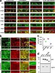
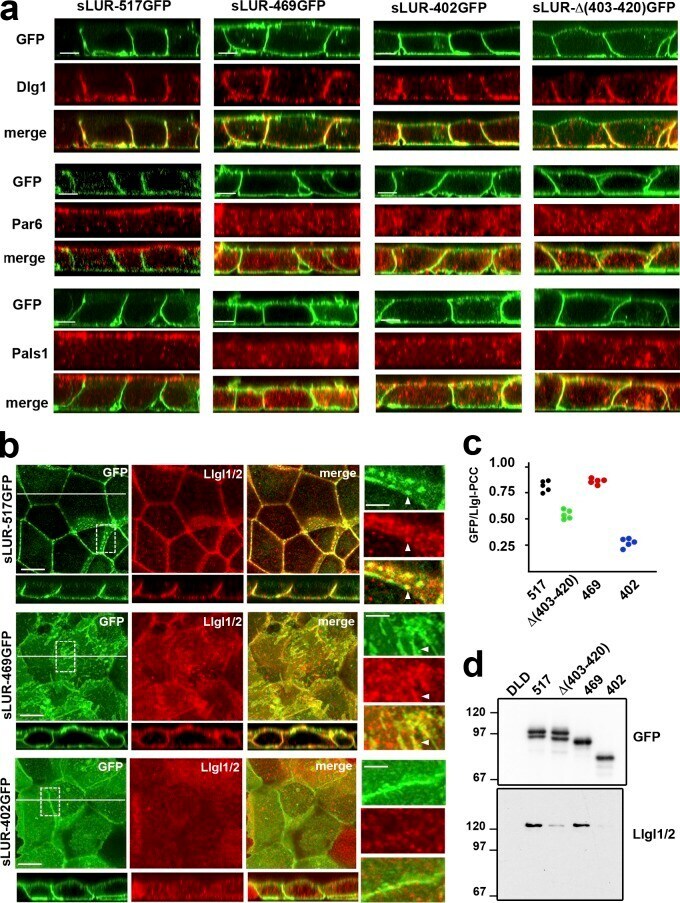
- Figure 10. Polarity markers in DLD20-2 cells expressing different sLUR mutants. (a) Representative z-sections of DLD20-2 cells expressing sLUR-517GFP, sLUR-469GFP, sLUR-402GFP, and sLUR-Delta(403-420). The cells were imaged for GFP fluorescence (green) in conjunction with immunostaining for Dlg1, Par6B, and Pals1 (all red). Bars, 10 um. Note that neither of the sLUR-517GFP mutants rescues ABP of DLD20-2 cells. (b) The cells were imaged for GFP (green) and for Llgl1/2 (red). x-y projections of three optical z slices spanning only 1.2 um of the apical cell regions are shown. The entire optical cross-sections of these cells along the white lines are at the bottom. Bars, 10 um. Dashed boxes show regions of higher magnifications (right). Note that some GFP and Llgl1/2 clusters are colocalized (arrowheads). (c) PCC values between green (GFP) and red (Llgl1/2) fluorescence of the random optical z slices of the images shown in b. (d) Western blot of the anti-GFP precipitates obtained from the confluent cultures of the control DLD cells and DLD20-2 cells expressing sLUR-517GFP mutants (marked as in Fig. 8, c and d ) probed for GFP and Llgl1/2.
- Submitted by
- Invitrogen Antibodies (provider)
- Main image
- Experimental details
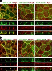
- Figure 5. Both PDZ domain-deficient GFP-sLUR and GFP-Lano rescue defects in polarity protein localization. (a and b) Projections of all optical slices of DLD20-2 cells expressing GFP-sLUR (a) and GFP-Lano (b). Cells were imaged for GFP fluorescence (green) in conjunction with anti-Llgl1/2, Pals1, or Par6B staining (all red). The corresponding x-z sections (with the same magnification) through the middle of each image are shown at the bottom. Bars, 10 um.

- Submitted by
- Invitrogen Antibodies (provider)
- Main image
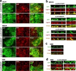
- Experimental details
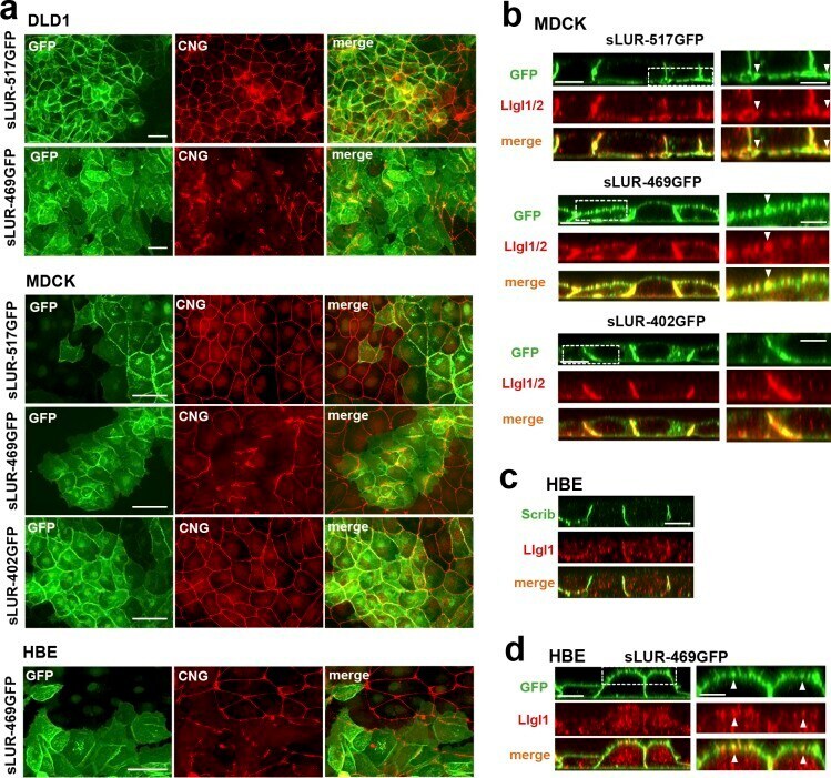
- Figure 9. Dominant-negative effect of the LAPSDb-deficient mutant. (a) Representative widefield images of DLD1, MDCK, and HBE cells expressing different sLUR mutants (indicated on the left) imaged for GFP (green) and CNG (red). The cells were cultured 48 h before staining. Note that the cells expressing the LAPSDb-deficient mutant, sLUR-469GFP, but not other mutants exhibit dramatic disintegration of TJs. Bars, 30 um. (b) Representative optical z-sections of MDCK cells expressing the full-length sLUR (sLUR-517GFP), and its mutants sLUR-469 GFP and sLUR-402GFP stained for Llgl1/2 (red). Note apical localization of Llgl1/2 in cells expressing sLUR-469GFP. Bars, 10 um. The boxed regions are zoomed on the right panel. Bars, 3 um. Note partial colocalization of the mutant and Llgl1/2 (arrowhead). (c) WT HBE cells stained for hScrib (Scrib) and Llgl1. Note the absence of both at the apical cortex. (d) HBE cells expressing sLUR-469GFP imaged for GFP and Llgl1. Note apical enrichment of both proteins. Bar, 10 um. The boxed area is zoomed on the right. Bar, 5 um.