Explore
 Validate
 Learn
Learn Western blot
 Immunocytochemistry
Antibody data
- Antibody Data
- Antigen structure
- References [14]
- Comments [0]
- Validations
- Immunocytochemistry [5]
- Other assay [15]
Submit
Validation data
Reference
Comment
Report error
- Product number
- PA1-025 - Provider product page

- Provider
- Invitrogen Antibodies
- Product name
- Cyclophilin A Polyclonal Antibody
- Antibody type
- Polyclonal
- Antigen
- Purifed from natural sources
- Description
- PA1-025 detects recombinant human cyclophilin A (CyPA), but does not detect endogenous levels of CyPA. PA1-025 also detects the CyPA protein from CHO cells. PA1-025 has been successfully used in Western blot and ICC/IF procedures. By Western blot, this antibody detects a prominent ~18 kDa protein representing recombinant human CyPA. The PA1-025 immunogen is recombinant human CyPA expressed in E. coli.
- Reactivity
- Human, Mouse, Hamster
- Host
- Rabbit
- Isotype
- IgG
- Vial size
- 400 μL
- Storage
- -20°C, Avoid Freeze/Thaw Cycles
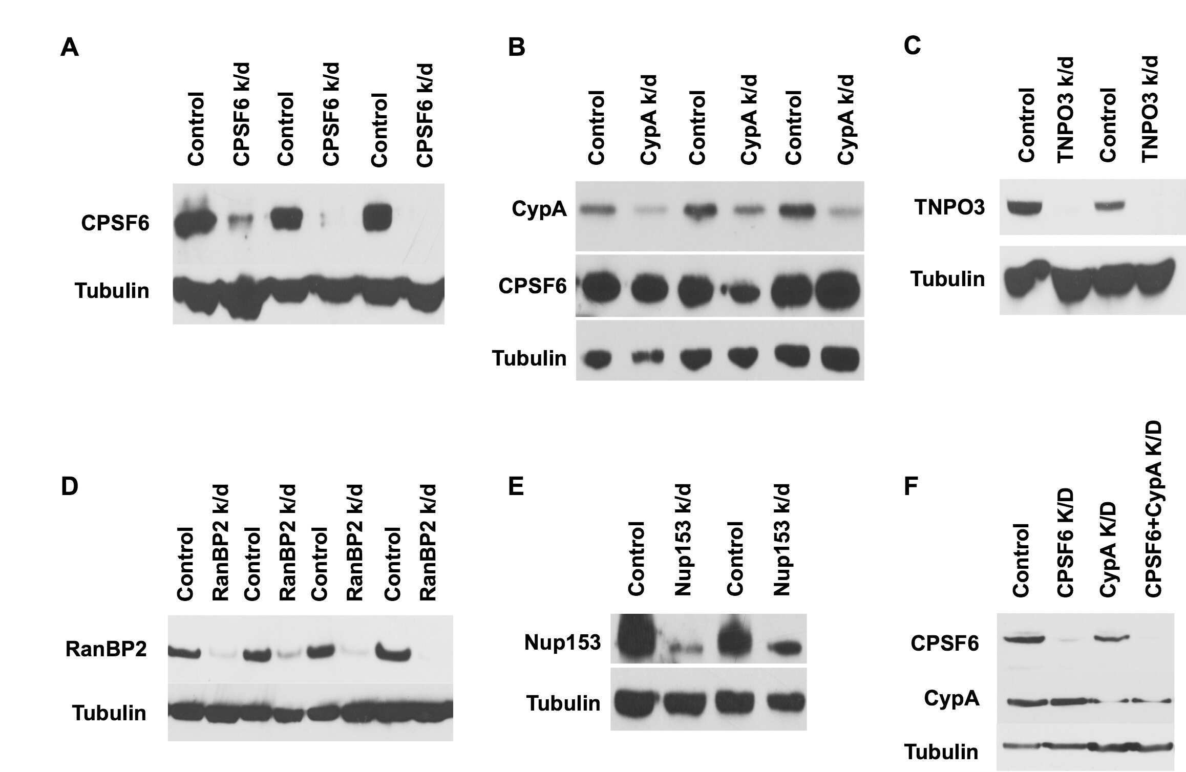
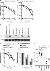
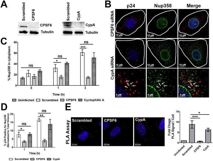
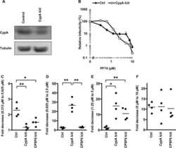
Submitted references KIF5B and Nup358 Cooperatively Mediate the Nuclear Import of HIV-1 during Infection.
Roles of Capsid-Interacting Host Factors in Multimodal Inhibition of HIV-1 by PF74.
In vivo functions of CPSF6 for HIV-1 as revealed by HIV-1 capsid evolution in HLA-B27-positive subjects.
Multiple sites in the N-terminal half of simian immunodeficiency virus capsid protein contribute to evasion from rhesus monkey TRIM5α-mediated restriction.
Evidence that mono-ADP-ribosylation of CtBP1/BARS regulates lipid storage.
Proteomic profiling identifies cyclooxygenase-2-independent global proteomic changes by celecoxib in colorectal cancer cells.
Cell surface expression of CD147/EMMPRIN is regulated by cyclophilin 60.
Cyclophilin interactions with incoming human immunodeficiency virus type 1 capsids with opposing effects on infectivity in human cells.
The characterization and hormonal regulation of kidney androgen-regulated protein (Kap)-luciferase transgenic mice.
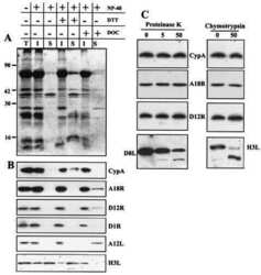
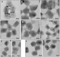
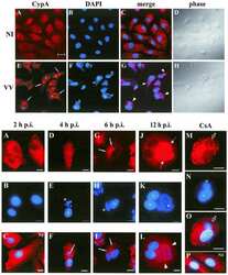
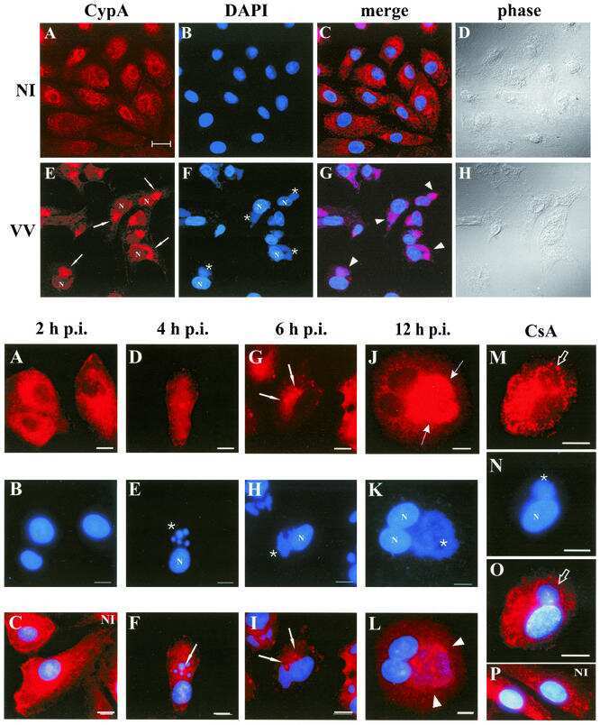
Redistribution of cyclophilin A to viral factories during vaccinia virus infection and its incorporation into mature particles.
Cyclophilin A peptidyl-prolyl isomerase activity promotes ZPR1 nuclear export.
Cholesteryl ester is transported from caveolae to internal membranes as part of a caveolin-annexin II lipid-protein complex.
Native recombinant cyclophilins A, B, and C degrade DNA independently of peptidylprolyl cis-trans-isomerase activity. Potential roles of cyclophilins in apoptosis.
A calcium-dependent nuclease from apoptotic rat thymocytes is homologous with cyclophilin. Recombinant cyclophilins A, B, and C have nuclease activity.
Dharan A, Talley S, Tripathi A, Mamede JI, Majetschak M, Hope TJ, Campbell EM
PLoS pathogens 2016 Jun;12(6):e1005700
PLoS pathogens 2016 Jun;12(6):e1005700
Roles of Capsid-Interacting Host Factors in Multimodal Inhibition of HIV-1 by PF74.
Saito A, Ferhadian D, Sowd GA, Serrao E, Shi J, Halambage UD, Teng S, Soto J, Siddiqui MA, Engelman AN, Aiken C, Yamashita M
Journal of virology 2016 Jun 15;90(12):5808-5823
Journal of virology 2016 Jun 15;90(12):5808-5823
In vivo functions of CPSF6 for HIV-1 as revealed by HIV-1 capsid evolution in HLA-B27-positive subjects.
Henning MS, Dubose BN, Burse MJ, Aiken C, Yamashita M
PLoS pathogens 2014 Jan;10(1):e1003868
PLoS pathogens 2014 Jan;10(1):e1003868
Multiple sites in the N-terminal half of simian immunodeficiency virus capsid protein contribute to evasion from rhesus monkey TRIM5α-mediated restriction.
Kono K, Song H, Yokoyama M, Sato H, Shioda T, Nakayama EE
Retrovirology 2010 Sep 8;7:72
Retrovirology 2010 Sep 8;7:72
Evidence that mono-ADP-ribosylation of CtBP1/BARS regulates lipid storage.
Bartz R, Seemann J, Zehmer JK, Serrero G, Chapman KD, Anderson RG, Liu P
Molecular biology of the cell 2007 Aug;18(8):3015-25
Molecular biology of the cell 2007 Aug;18(8):3015-25
Proteomic profiling identifies cyclooxygenase-2-independent global proteomic changes by celecoxib in colorectal cancer cells.
Lou J, Fatima N, Xiao Z, Stauffer S, Smythers G, Greenwald P, Ali IU
Cancer epidemiology, biomarkers & prevention : a publication of the American Association for Cancer Research, cosponsored by the American Society of Preventive Oncology 2006 Sep;15(9):1598-606
Cancer epidemiology, biomarkers & prevention : a publication of the American Association for Cancer Research, cosponsored by the American Society of Preventive Oncology 2006 Sep;15(9):1598-606
Cell surface expression of CD147/EMMPRIN is regulated by cyclophilin 60.
Pushkarsky T, Yurchenko V, Vanpouille C, Brichacek B, Vaisman I, Hatakeyama S, Nakayama KI, Sherry B, Bukrinsky MI
The Journal of biological chemistry 2005 Jul 29;280(30):27866-71
The Journal of biological chemistry 2005 Jul 29;280(30):27866-71
Cyclophilin interactions with incoming human immunodeficiency virus type 1 capsids with opposing effects on infectivity in human cells.
Hatziioannou T, Perez-Caballero D, Cowan S, Bieniasz PD
Journal of virology 2005 Jan;79(1):176-83
Journal of virology 2005 Jan;79(1):176-83
The characterization and hormonal regulation of kidney androgen-regulated protein (Kap)-luciferase transgenic mice.
Malstrom SE, Tornavaca O, Meseguer A, Purchio AF, West DB
Toxicological sciences : an official journal of the Society of Toxicology 2004 Jun;79(2):266-77
Toxicological sciences : an official journal of the Society of Toxicology 2004 Jun;79(2):266-77
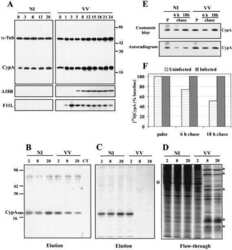
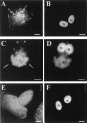
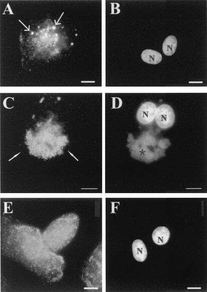
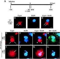
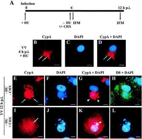
Redistribution of cyclophilin A to viral factories during vaccinia virus infection and its incorporation into mature particles.
Castro AP, Carvalho TM, Moussatché N, Damaso CR
Journal of virology 2003 Aug;77(16):9052-68
Journal of virology 2003 Aug;77(16):9052-68
Cyclophilin A peptidyl-prolyl isomerase activity promotes ZPR1 nuclear export.
Ansari H, Greco G, Luban J
Molecular and cellular biology 2002 Oct;22(20):6993-7003
Molecular and cellular biology 2002 Oct;22(20):6993-7003
Cholesteryl ester is transported from caveolae to internal membranes as part of a caveolin-annexin II lipid-protein complex.
Uittenbogaard A, Everson WV, Matveev SV, Smart EJ
The Journal of biological chemistry 2002 Feb 15;277(7):4925-31
The Journal of biological chemistry 2002 Feb 15;277(7):4925-31
Native recombinant cyclophilins A, B, and C degrade DNA independently of peptidylprolyl cis-trans-isomerase activity. Potential roles of cyclophilins in apoptosis.
Montague JW, Hughes FM Jr, Cidlowski JA
The Journal of biological chemistry 1997 Mar 7;272(10):6677-84
The Journal of biological chemistry 1997 Mar 7;272(10):6677-84
A calcium-dependent nuclease from apoptotic rat thymocytes is homologous with cyclophilin. Recombinant cyclophilins A, B, and C have nuclease activity.
Montague JW, Gaido ML, Frye C, Cidlowski JA
The Journal of biological chemistry 1994 Jul 22;269(29):18877-80
The Journal of biological chemistry 1994 Jul 22;269(29):18877-80
No comments: Submit comment
Supportive validation
- Submitted by
- Invitrogen Antibodies (provider)
- Main image
- Experimental details
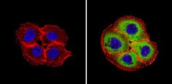
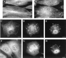
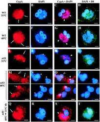
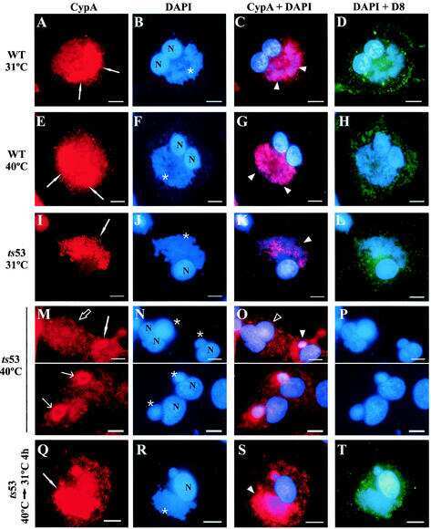
- Immunofluorescent analysis of Cyclophilin A (green) showing staining in the in the cytoplasm of A431 cells (right) compared to a negative control without primary antibody (left). Formalin-fixed cells were permeabilized with 0.1% Triton X-100 in TBS for 5-10 minutes and blocked with 3% BSA-PBS for 30 minutes at room temperature. Cells were probed with a Cyclophilin A monoclonal antibody (Product # PA1-025) in 3% BSA-PBS at a dilution of 1:200 and incubated overnight at 4ºC in a humidified chamber. Cells were washed with PBST and incubated with a DyLight-conjugated secondary antibody in PBS at room temperature in the dark. F-actin (red) was stained with a fluorescent red phalloidin and nuclei (blue) were stained with Hoechst or DAPI. Images were taken at a magnification of 60x.

- Submitted by
- Invitrogen Antibodies (provider)
- Main image

- Experimental details
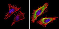
- Immunofluorescent analysis of Cyclophilin A (green) showing staining in the in the cytoplasm of Hela cells (right) compared to a negative control without primary antibody (left). Formalin-fixed cells were permeabilized with 0.1% Triton X-100 in TBS for 5-10 minutes and blocked with 3% BSA-PBS for 30 minutes at room temperature. Cells were probed with a Cyclophilin A polyclonal antibody (Product # PA1-025) in 3% BSA-PBS at a dilution of 1:200 and incubated overnight at 4ºC in a humidified chamber. Cells were washed with PBST and incubated with a DyLight-conjugated secondary antibody in PBS at room temperature in the dark. F-actin (red) was stained with a fluorescent red phalloidin and nuclei (blue) were stained with Hoechst or DAPI. Images were taken at a magnification of 60x.

- Submitted by
- Invitrogen Antibodies (provider)
- Main image
- Experimental details
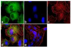
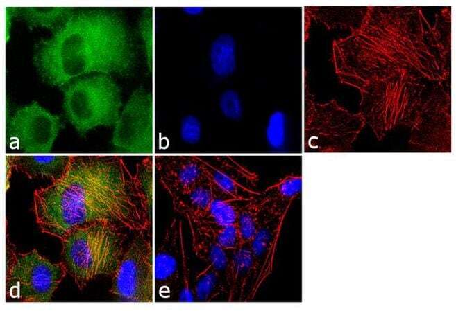
- Immunofluorescence analysis of Cyclophilin A was performed using 70% confluent log phase HeLa cells. The cells were fixed with 4% paraformaldehyde for 10 minutes, permeabilized with 0.1% Triton™ X-100 for 10 minutes, and blocked with 1% BSA for 1 hour at room temperature. The cells were labeled with Cyclophilin A Rabbit Polyclonal Antibody (Product # PA1-025) at 1:250 dilution in 0.1% BSA and incubated for 3 hours at room temperature and then labeled with Goat anti-Rabbit IgG (H+L) Superclonal™ Secondary Antibody, Alexa Fluor® 488 conjugate (Product # A27034) at a dilution of 1:2000 for 45 minutes at room temperature (Panel a: green). Nuclei (Panel b: blue) were stained with SlowFade® Gold Antifade Mountant with DAPI (Product # S36938). F-actin (Panel c: red) was stained with Rhodamine Phalloidin (Product # R415, 1:300). Panel d represents the merged image showing cytoplasmic localization. Panel e shows the no primary antibody control. The images were captured at 60X magnification.
- Submitted by
- Invitrogen Antibodies (provider)
- Main image
- Experimental details
- Immunofluorescent analysis of Cyclophilin A (green) showing staining in the in the cytoplasm of Hela cells (right) compared to a negative control without primary antibody (left). Formalin-fixed cells were permeabilized with 0.1% Triton X-100 in TBS for 5-10 minutes and blocked with 3% BSA-PBS for 30 minutes at room temperature. Cells were probed with a Cyclophilin A polyclonal antibody (Product # PA1-025) in 3% BSA-PBS at a dilution of 1:200 and incubated overnight at 4ºC in a humidified chamber. Cells were washed with PBST and incubated with a DyLight-conjugated secondary antibody in PBS at room temperature in the dark. F-actin (red) was stained with a fluorescent red phalloidin and nuclei (blue) were stained with Hoechst or DAPI. Images were taken at a magnification of 60x.

- Submitted by
- Invitrogen Antibodies (provider)
- Main image

- Experimental details
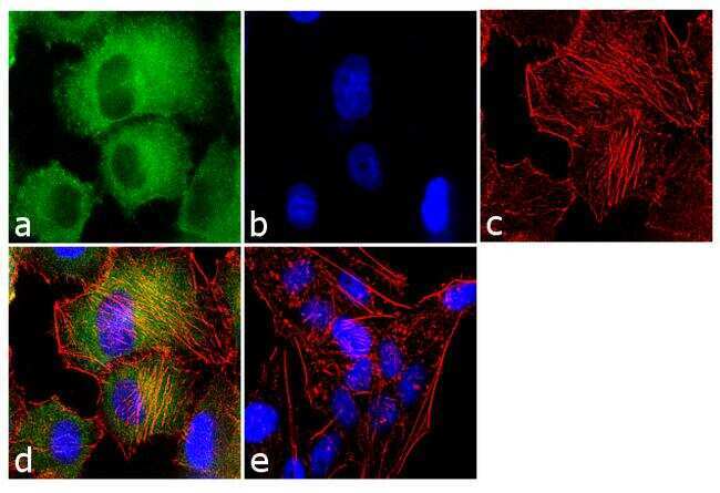
- Immunofluorescence analysis of Cyclophilin A was performed using 70% confluent log phase HeLa cells. The cells were fixed with 4% paraformaldehyde for 10 minutes, permeabilized with 0.1% Triton™ X-100 for 10 minutes, and blocked with 1% BSA for 1 hour at room temperature. The cells were labeled with Cyclophilin A Rabbit Polyclonal Antibody (Product # PA1-025) at 1:250 dilution in 0.1% BSA and incubated for 3 hours at room temperature and then labeled with Goat anti-Rabbit IgG (Heavy Chain) Superclonal™ Secondary Antibody, Alexa Fluor® 488 conjugate (Product # A27034) at a dilution of 1:2000 for 45 minutes at room temperature (Panel a: green). Nuclei (Panel b: blue) were stained with SlowFade® Gold Antifade Mountant with DAPI (Product # S36938). F-actin (Panel c: red) was stained with Rhodamine Phalloidin (Product # R415, 1:300). Panel d represents the merged image showing cytoplasmic localization. Panel e shows the no primary antibody control. The images were captured at 60X magnification.
Supportive validation
- Submitted by
- Invitrogen Antibodies (provider)
- Main image

- Experimental details
- NULL
- Submitted by
- Invitrogen Antibodies (provider)
- Main image
- Experimental details
- NULL
- Submitted by
- Invitrogen Antibodies (provider)
- Main image
- Experimental details
- NULL
- Submitted by
- Invitrogen Antibodies (provider)
- Main image
- Experimental details
- NULL

- Submitted by
- Invitrogen Antibodies (provider)
- Main image
- Experimental details
- NULL
- Submitted by
- Invitrogen Antibodies (provider)
- Main image
- Experimental details
- NULL
- Submitted by
- Invitrogen Antibodies (provider)
- Main image
- Experimental details
- NULL
- Submitted by
- Invitrogen Antibodies (provider)
- Main image

- Experimental details
- NULL
- Submitted by
- Invitrogen Antibodies (provider)
- Main image

- Experimental details
- NULL
- Submitted by
- Invitrogen Antibodies (provider)
- Main image
- Experimental details
- NULL

- Submitted by
- Invitrogen Antibodies (provider)
- Main image
- Experimental details
- NULL

- Submitted by
- Invitrogen Antibodies (provider)
- Main image
- Experimental details
- NULL
- Submitted by
- Invitrogen Antibodies (provider)
- Main image
- Experimental details
- NULL

- Submitted by
- Invitrogen Antibodies (provider)
- Main image

- Experimental details
- NULL
- Submitted by
- Invitrogen Antibodies (provider)
- Main image
- Experimental details
- NULL