Explore
 Validate
 Learn
LearnMA1-10192
antibody from Invitrogen Antibodies
Targeting: CDH1
CD324, UVO, uvomorulin
 Western blot
 Immunocytochemistry Immunoprecipitation Immunohistochemistry Flow cytometry Other assay
Antibody data
- Antibody Data
- Antigen structure
- References [3]
- Comments [0]
- Validations
- Immunocytochemistry [1]
- Other assay [2]
Submit
Validation data
Reference
Comment
Report error
- Product number
- MA1-10192 - Provider product page

- Provider
- Invitrogen Antibodies
- Product name
- E-cadherin Monoclonal Antibody (67A4)
- Antibody type
- Monoclonal
- Antigen
- Other
- Description
- This antibody recognizes an extracellular epitope of CD324 / E-cadherin, an approximately 100 kDa epithelial cell adhesion molecule, whose detection is important for determination of invasive potential of epithelial neoplasms.
- Reactivity
- Human
- Host
- Mouse
- Isotype
- IgG
- Antibody clone number
- 67A4
- Vial size
- 100 µg
- Concentration
- 1.0 mg/mL
- Storage
- 4°C, do not freeze
Submitted references The use of patient-derived breast tissue explants to study macrophage polarization and the effects of environmental chemical exposure.
mRNA and miRNA expression profiles in an ectoderm-biased substate of human pluripotent stem cells.
NKX2-5 regulates human cardiomyogenesis via a HEY2 dependent transcriptional network.
Gregory KJ, Morin SM, Kubosiak A, Ser-Dolansky J, Schalet BJ, Jerry DJ, Schneider SS
Immunology and cell biology 2020 Nov;98(10):883-896
Immunology and cell biology 2020 Nov;98(10):883-896
mRNA and miRNA expression profiles in an ectoderm-biased substate of human pluripotent stem cells.
Mawaribuchi S, Aiki Y, Ikeda N, Ito Y
Scientific reports 2019 Aug 15;9(1):11910
Scientific reports 2019 Aug 15;9(1):11910
NKX2-5 regulates human cardiomyogenesis via a HEY2 dependent transcriptional network.
Anderson DJ, Kaplan DI, Bell KM, Koutsis K, Haynes JM, Mills RJ, Phelan DG, Qian EL, Leitoguinho AR, Arasaratnam D, Labonne T, Ng ES, Davis RP, Casini S, Passier R, Hudson JE, Porrello ER, Costa MW, Rafii A, Curl CL, Delbridge LM, Harvey RP, Oshlack A, Cheung MM, Mummery CL, Petrou S, Elefanty AG, Stanley EG, Elliott DA
Nature communications 2018 Apr 10;9(1):1373
Nature communications 2018 Apr 10;9(1):1373
No comments: Submit comment
Supportive validation
- Submitted by
- Invitrogen Antibodies (provider)
- Main image

- Experimental details
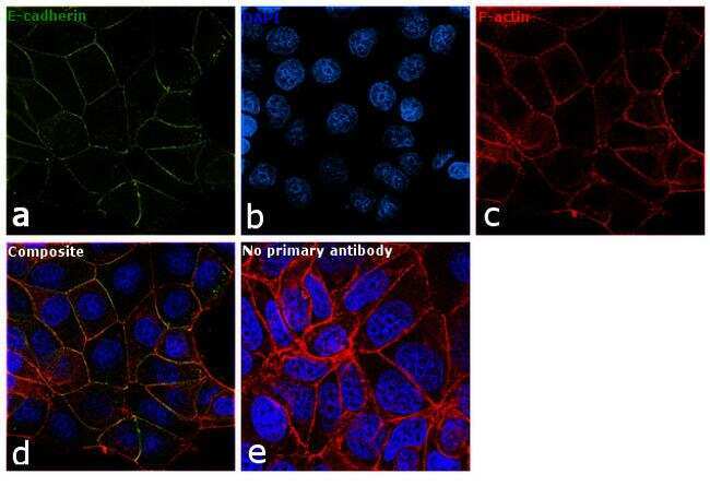
- Immunofluorescence analysis of E-cadherin was performed using 90% confluent log phase MCF7 cells. The cells were fixed with 4% paraformaldehyde for 10 minutes, permeabilized with 0.1% Triton™ X-100 for 15 minutes, and blocked with 1% BSA for 1 hour at room temperature. The cells were labeled with E-cadherin Monoclonal Antibody (67A4) (Product # MA1-10192) at 5 µg/mL in 0.1% BSA, incubated at 4 degree Celsius overnight and then labeled with Goat anti-Mouse IgG (H+L) Superclonal™ Secondary Antibody, Alexa Fluor® 488 conjugate (Product # A28175) at a dilution of 1:2000 for 45 minutes at room temperature (Panel a: green). Nuclei (Panel b: blue) were stained with SlowFade® Gold Antifade Mountant with DAPI (Product # S36938). F-actin (Panel c: red) was stained with Rhodamine Phalloidin (Product # R415, 1:300). Panel d represents the merged image showing plasma membrane localization. Panel e represents control cells with no primary antibody to assess background. The images were captured at 60X magnification.
Supportive validation
- Submitted by
- Invitrogen Antibodies (provider)
- Main image
- Experimental details
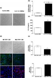
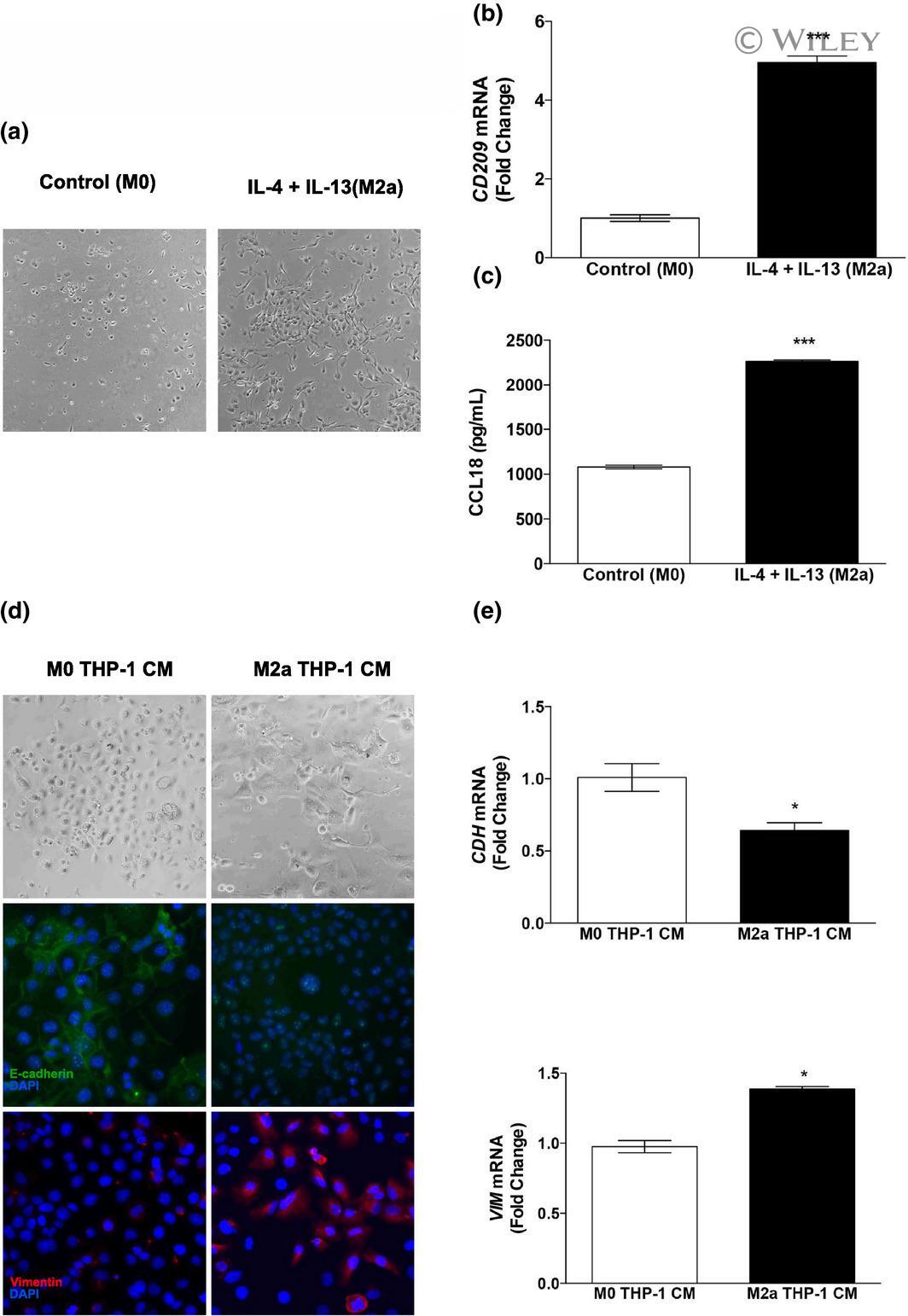
- 4 Figure In vitro polarization of THP-1 cells toward the M(IL-4 + IL-13) phenotype and the effects of THP-1 CM on EMT in MCF10A cells. THP-1 monocytes were treated with a combination of PMA, IL-4 and IL-13 and (a) images were captured to illustrate the morphological changes in response to cytokine treatment. (b) CD209 mRNA was quantified by real-time PCR and (c) CCL18 secretion in cell culture medium was measured by ELISA in technical duplicates. MCF10A cells treated with THP-1 CM were grown to 50% confluence and (d) images were captured to illustrate the morphological changes in response to THP-1 CM treatment. The cells were then immunostained with either anti-E-cadherin or anti-vimentin antibodies and counterstained with DAPI. (e) RNA was harvested from THP-1 CM-treated cells and real-time PCR analysis of CDH and VIM was carried out. Data within each bar represent triplicate treatments, are presented as mean +- s.e.m. and are expressed as fold change with respect to controls. * P < 0.05, *** P < 0.001 (significantly different from indicated data set using a Student's t -test). All images were captured at 400x magnification. CM, conditioned medium; DAPI, 4',6-diamidino-2-phenylindole; EMT, epithelial-to-mesenchymal transition; IL, interleukin; mRNA, messenger RNA; PMA, phorbol 12-myristate 13-acetate.
- Submitted by
- Invitrogen Antibodies (provider)
- Main image
- Experimental details
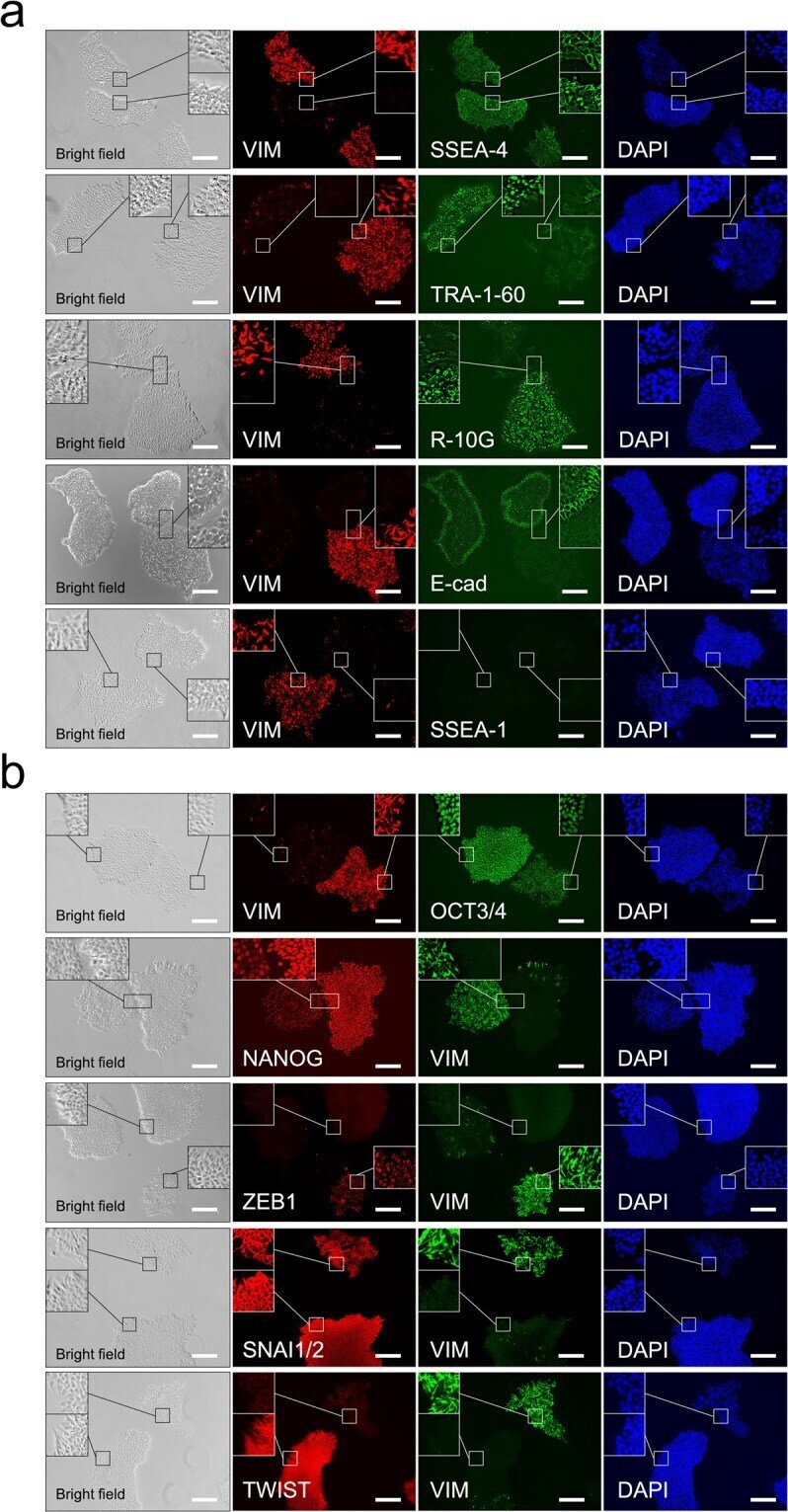
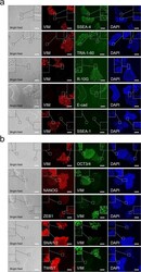
- Figure 3 Immunohistochemical analysis of pluripotency, differentiation, and epithelial-to-mesenchymal transition markers in H9 cells cultured on Matrigel. ( a ) Immunohistochemistry with no permeabilisation using antibodies for vimentin (VIM), SSEA-4, TRA-1-60, R-10G, E-cadherin (E-cad), and SSEA-1. ( b ) Immunohistochemistry with permeabilisation using antibodies for VIM, OCT3/4, NANOG, ZEB1, SNAI1/2, and TWIST. Nuclei were counterstained with DAPI. The inset shows a 3 x enlargement of the figure. The scale bar represents 200 um.