Explore
 Validate
 Learn
Learn Western blot
 Immunohistochemistry
Antibody data
- Antibody Data
- Antigen structure
- References [2]
- Comments [0]
- Validations
- Immunohistochemistry [2]
- Flow cytometry [8]
Submit
Validation data
Reference
Comment
Report error
- Product number
- MA5-16086 - Provider product page

- Provider
- Invitrogen Antibodies
- Product name
- Tenascin C Monoclonal Antibody (4C8MS)
- Antibody type
- Monoclonal
- Antigen
- Recombinant full-length protein
- Reactivity
- Human, Mouse, Rat, Feline
- Host
- Mouse
- Isotype
- IgG
- Antibody clone number
- 4C8MS
- Vial size
- 100 μL
- Concentration
- 1.0 mg/mL
- Storage
- Store at 4°C short term. For long term storage, store at -20°C, avoiding freeze/thaw cycles.
Submitted references The Effects of Cold Water Immersion and Active Recovery on Molecular Factors That Regulate Growth and Remodeling of Skeletal Muscle After Resistance Exercise.
Targeted disruption of the iNOS gene improves adipose tissue inflammation and fibrosis in leptin-deficient ob/ob mice: role of tenascin C.
Peake JM, Markworth JF, Cumming KT, Aas SN, Roberts LA, Raastad T, Cameron-Smith D, Figueiredo VC
Frontiers in physiology 2020;11:737
Frontiers in physiology 2020;11:737
Targeted disruption of the iNOS gene improves adipose tissue inflammation and fibrosis in leptin-deficient ob/ob mice: role of tenascin C.
Becerril S, Rodríguez A, Catalán V, Méndez-Giménez L, Ramírez B, Sáinz N, Llorente M, Unamuno X, Gómez-Ambrosi J, Frühbeck G
International journal of obesity (2005) 2018 Aug;42(8):1458-1470
International journal of obesity (2005) 2018 Aug;42(8):1458-1470
No comments: Submit comment
Supportive validation
- Submitted by
- Invitrogen Antibodies (provider)
- Main image
- Experimental details
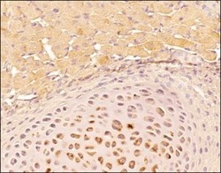
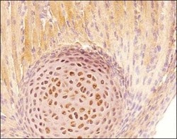
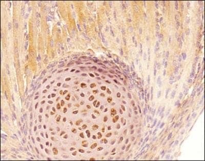
- Immunohistochemical analysis of Tenascin C in formalin fixed paraffin embedded tissue section of mouse bone-tendon. Samples were incubated in Tenascin C monoclonal antibody (Product # MA5-16086) using a dilution of 1:25. The signal was detected using HRP-DAB detection method which followed counterstaining using hematoxylin. The antibody generated a very specific cytoplasmic, membrane and extra-cellular signal in tendon fibroblasts, osteoblasts, osteoclasts, and some bone marrow cells. The mineralized areas were largely negative for the staining.

- Submitted by
- Invitrogen Antibodies (provider)
- Main image
- Experimental details
- Immunohistochemical analysis of Tenascin C in formalin fixed paraffin embedded tissue section of mouse bone-tendon. Samples were incubated in Tenascin C monoclonal antibody (Product # MA5-16086) using a dilution of 1:25. The signal was detected using HRP-DAB detection method which followed counterstaining using hematoxylin. The antibody generated a very specific cytoplasmic, membrane and extra-cellular signal in tendon fibroblasts, osteoblasts, osteoclasts, and some bone marrow cells. The mineralized areas were largely negative for the staining.
Supportive validation
- Submitted by
- Invitrogen Antibodies (provider)
- Main image
- Experimental details
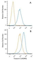
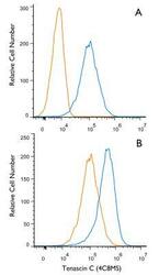
- Flow cytometry of Tenascin C in U87MG Cells. Samples were incubated in Tenascin C monoclonal antibody (Product # MA5-16086) and a matched isotype control using a dilution of 1 µg/mL for 30 minutes at room temperature followed by mouse F(ab)2 IgG (H+L) APC-conjugated secondary antibody. Figure A: Antibody (blue) and a matched isotype control NBP1-97005 (orange). Cells were fixed with 4% paraformaldehyde, following fixation, cells were permeabilized with 0.1% saponin. Figure B: Untreated cells (orange) or treated cells with 3 µM Monensin (blue). Cells were fixed with 4% paraformaldehyde, following fixation, cells were permeabilized with 0.1% saponin.
- Submitted by
- Invitrogen Antibodies (provider)
- Main image
- Experimental details
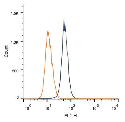
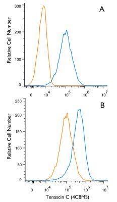
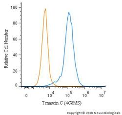
- Flow cytometry of Tenascin C in SK-MEL-28 cells. Samples were incubated in Tenascin C monoclonal antibody (Product # MA5-16086) and a matched isotype control using a dilution of 2.5 µg/mL for 30 minutes at room temperature followed by mouse F(ab)2 IgG (H+L) APC-conjugated secondary antibody. Cells were fixed with 4% PFA and then permeablized with 0.1% saponin.
- Submitted by
- Invitrogen Antibodies (provider)
- Main image

- Experimental details
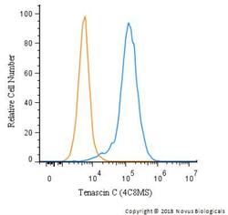
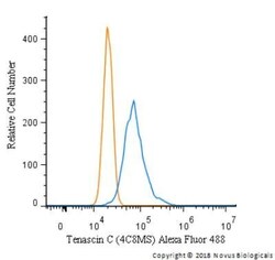
- Flow cytometry of Tenascin C in SK-MEL-28 cells (blue) and a matched isotype control (orange). Samples were incubated in Tenascin C monoclonal antibody (Product # MA5-16086) using a dilution of 5 µg/mL for 30 minutes at room temperature. Cells were fixed with 4% PFA and then permeabilized with 0.1% saponin. Both antibodies were conjugated to Alexa Fluor 488.

- Submitted by
- Invitrogen Antibodies (provider)
- Main image
- Experimental details
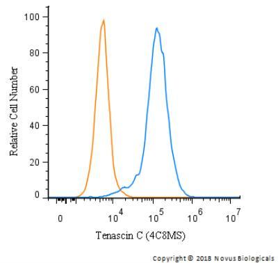
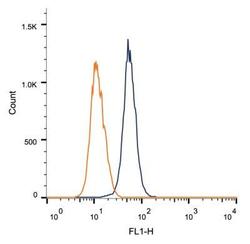
- Flow cytometry of Tenascin C in 1 x 10^6 MCF-7 cells. Samples were incubated in Tenascin C monoclonal antibody (Product # MA5-16086) using a dilution of 1 µg/1x10^6 cells. Antibody (dark blue). Isotype control shown in orange.

- Submitted by
- Invitrogen Antibodies (provider)
- Main image
- Experimental details
- Flow cytometry of Tenascin C in U87MG Cells. Samples were incubated in Tenascin C monoclonal antibody (Product # MA5-16086) and a matched isotype control using a dilution of 1 µg/mL for 30 minutes at room temperature followed by mouse F(ab)2 IgG (H+L) APC-conjugated secondary antibody. Figure A: Antibody (blue) and a matched isotype control NBP1-97005 (orange). Cells were fixed with 4% paraformaldehyde, following fixation, cells were permeabilized with 0.1% saponin. Figure B: Untreated cells (orange) or treated cells with 3 µM Monensin (blue). Cells were fixed with 4% paraformaldehyde, following fixation, cells were permeabilized with 0.1% saponin.

- Submitted by
- Invitrogen Antibodies (provider)
- Main image
- Experimental details
- Flow cytometry of Tenascin C in SK-MEL-28 cells. Samples were incubated in Tenascin C monoclonal antibody (Product # MA5-16086) and a matched isotype control using a dilution of 2.5 µg/mL for 30 minutes at room temperature followed by mouse F(ab)2 IgG (H+L) APC-conjugated secondary antibody. Cells were fixed with 4% PFA and then permeablized with 0.1% saponin.

- Submitted by
- Invitrogen Antibodies (provider)
- Main image
- Experimental details
- Flow cytometry of Tenascin C in SK-MEL-28 cells (blue) and a matched isotype control (orange). Samples were incubated in Tenascin C monoclonal antibody (Product # MA5-16086) using a dilution of 5 µg/mL for 30 minutes at room temperature. Cells were fixed with 4% PFA and then permeabilized with 0.1% saponin. Both antibodies were conjugated to Alexa Fluor 488.

- Submitted by
- Invitrogen Antibodies (provider)
- Main image

- Experimental details
- Flow cytometry of Tenascin C in 1 x 10^6 MCF-7 cells. Samples were incubated in Tenascin C monoclonal antibody (Product # MA5-16086) using a dilution of 1 µg/1x10^6 cells. Antibody (dark blue). Isotype control shown in orange.