 Explore
 Validate
 Learn
Learn Western blot
 Immunocytochemistry
Antibody data
- Antibody Data
- Antigen structure
- References [8]
- Comments [0]
- Validations
- Immunocytochemistry [1]
Submit
Validation data
Reference
Comment
Report error
- Product number
- MAB2138 - Provider product page

- Provider
- R&D Systems
- Product name
- Human/Mouse Tenascin C Antibody
- Antibody type
- Monoclonal
- Description
- Protein A or G purified from hybridoma culture supernatant. Detect human and mouse Tenascin C in Western blots.
- Reactivity
- Human, Mouse
- Host
- Rat
- Conjugate
- Unconjugated
- Isotype
- IgG
- Antibody clone number
- 578
- Vial size
- 100 ug
- Concentration
- LYOPH
- Storage
- Use a manual defrost freezer and avoid repeated freeze-thaw cycles. 12 months from date of receipt, -20 to -70 °C as supplied. 1 month, 2 to 8 °C under sterile conditions after reconstitution. 6 months, -20 to -70 °C under sterile conditions after reconstitution.
Submitted references Combined CSL and p53 downregulation promotes cancer-associated fibroblast activation.
Temporal expression of growth factors triggered by epiregulin regulates inflammation development.
The missense mutation p.R1303Q in type XVII collagen underlies junctional epidermolysis bullosa resembling Kindler syndrome.
Melanoma cell invasiveness is promoted at least in part by the epidermal growth factor-like repeats of tenascin-C.
Mechanisms of fibroblast cell therapy for dystrophic epidermolysis bullosa: high stability of collagen VII favors long-term skin integrity.
ELR-negative CXC chemokine CXCL11 (IP-9/I-TAC) facilitates dermal and epidermal maturation during wound repair.
A hypomorphic mouse model of dystrophic epidermolysis bullosa reveals mechanisms of disease and response to fibroblast therapy.
Essential role of Smad3 in infarct healing and in the pathogenesis of cardiac remodeling.
Procopio MG, Laszlo C, Al Labban D, Kim DE, Bordignon P, Jo SH, Goruppi S, Menietti E, Ostano P, Ala U, Provero P, Hoetzenecker W, Neel V, Kilarski WW, Swartz MA, Brisken C, Lefort K, Dotto GP
Nature cell biology 2015 Sep;17(9):1193-204
Nature cell biology 2015 Sep;17(9):1193-204
Temporal expression of growth factors triggered by epiregulin regulates inflammation development.
Harada M, Kamimura D, Arima Y, Kohsaka H, Nakatsuji Y, Nishida M, Atsumi T, Meng J, Bando H, Singh R, Sabharwal L, Jiang JJ, Kumai N, Miyasaka N, Sakoda S, Yamauchi-Takihara K, Ogura H, Hirano T, Murakami M
Journal of immunology (Baltimore, Md. : 1950) 2015 Feb 1;194(3):1039-46
Journal of immunology (Baltimore, Md. : 1950) 2015 Feb 1;194(3):1039-46
The missense mutation p.R1303Q in type XVII collagen underlies junctional epidermolysis bullosa resembling Kindler syndrome.
Has C, Kiritsi D, Mellerio JE, Franzke CW, Wedgeworth E, Tantcheva-Poor I, Kernland-Lang K, Itin P, Simpson MA, Dopping-Hepenstal PJ, Fujimoto W, McGrath JA, Bruckner-Tuderman L
The Journal of investigative dermatology 2014 Mar;134(3):845-849
The Journal of investigative dermatology 2014 Mar;134(3):845-849
Melanoma cell invasiveness is promoted at least in part by the epidermal growth factor-like repeats of tenascin-C.
Grahovac J, Becker D, Wells A
The Journal of investigative dermatology 2013 Jan;133(1):210-20
The Journal of investigative dermatology 2013 Jan;133(1):210-20
Mechanisms of fibroblast cell therapy for dystrophic epidermolysis bullosa: high stability of collagen VII favors long-term skin integrity.
Kern JS, Loeckermann S, Fritsch A, Hausser I, Roth W, Magin TM, Mack C, Müller ML, Paul O, Ruther P, Bruckner-Tuderman L
Molecular therapy : the journal of the American Society of Gene Therapy 2009 Sep;17(9):1605-15
Molecular therapy : the journal of the American Society of Gene Therapy 2009 Sep;17(9):1605-15
ELR-negative CXC chemokine CXCL11 (IP-9/I-TAC) facilitates dermal and epidermal maturation during wound repair.
Yates CC, Whaley D, Y-Chen A, Kulesekaran P, Hebda PA, Wells A
The American journal of pathology 2008 Sep;173(3):643-52
The American journal of pathology 2008 Sep;173(3):643-52
A hypomorphic mouse model of dystrophic epidermolysis bullosa reveals mechanisms of disease and response to fibroblast therapy.
Fritsch A, Loeckermann S, Kern JS, Braun A, Bösl MR, Bley TA, Schumann H, von Elverfeldt D, Paul D, Erlacher M, Berens von Rautenfeld D, Hausser I, Fässler R, Bruckner-Tuderman L
The Journal of clinical investigation 2008 May;118(5):1669-79
The Journal of clinical investigation 2008 May;118(5):1669-79
Essential role of Smad3 in infarct healing and in the pathogenesis of cardiac remodeling.
Bujak M, Ren G, Kweon HJ, Dobaczewski M, Reddy A, Taffet G, Wang XF, Frangogiannis NG
Circulation 2007 Nov 6;116(19):2127-38
Circulation 2007 Nov 6;116(19):2127-38
No comments: Submit comment
Supportive validation
- Submitted by
- R&D Systems (provider)
- Main image
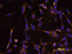
- Experimental details
- Tenascin C in U-118 MG Human Cell Line. Tenascin C was detected in immersion fixed U-118 MG human glioblastoma/astrocytoma cell line using Rat Anti-Human/Mouse Tenascin C Monoclonal Antibody (Catalog # MAB2138) at 10 µg/mL for 3 hours at room temperature. Cells were stained using the NorthernLights™ 557-conjugated Anti-Rat IgG Secondary Antibody (yellow; Catalog # NL013) and counterstained with DAPI (blue). View our protocol for Fluorescent ICC Staining of Cells on Coverslips.
