Explore
 Validate
 Learn
Learn Immunocytochemistry
Antibody data
- Antibody Data
- Antigen structure
- References [4]
- Comments [0]
- Validations
- Immunocytochemistry [1]
- Flow cytometry [1]
- Other assay [4]
Submit
Validation data
Reference
Comment
Report error
- Product number
- 53-9159-82 - Provider product page

- Provider
- Invitrogen Antibodies
- Product name
- Desmoglein 2 Monoclonal Antibody (CSTEM28), Alexa Fluor™ 488, eBioscience™
- Antibody type
- Monoclonal
- Antigen
- Other
- Description
- This monoclonal antibody CSTEM28 reacts with human desmoglein 2 (DSG2), a type 1 transmembrane protein belonging to the cadherin family. DSG2 is expressed by epithelial cells, cardiomyocytes, endothelial progenitors, and CD34+CD45dim hematopoietic stem and progenitor cells. This adhesion molecule forms cell-cell adhesion complexes called desmosomes that provide tensile strength to tissues that experience mechanical stress. Other than its role in desmosome formation, DSG2 plays a role in barrier integrity, adhesion, migration, cell apoptosis, proliferation, vasculogenic mimicry, and intracellular signaling. DSG2-expressing endothelial progenitor cells are pro-angiogenic, suggesting a role in vasculature formation. On hematopoietic stem and progenitor cells, DSG2 expression is highest on CD38-CD90+ multi-potent cells. DSG2 expression is progressively decreased as a function of differentiation, with DSG2 expression absent on mature lymphocytes. DSG2 also serves as a receptor for adenoviruses and can be shed by mucosal barriers in the intestine. This CSTEM28 antibody has been tested by flow cytometric analysis of 2102ep cell line. This has also been tested by immunocytochemistry of fixed and permeabilized cells and can be used at less than or equal to 10 µg/mL. It is recommended the antibody be carefully titrated for optimal performance in the assay of interest. Filtration: 0.2 µm post-manufacturing filtered.
- Reactivity
- Human
- Host
- Mouse
- Conjugate
- Green dye
- Isotype
- IgG
- Antibody clone number
- CSTEM28
- Vial size
- 100 µg
- Concentration
- 0.5 mg/mL
- Storage
- 4° C, store in dark, DO NOT FREEZE!
Submitted references External Beam Radiation Therapy and Enadenotucirev: Inhibition of the DDR and Mechanisms of Radiation-Mediated Virus Increase.
Comparative Analysis of Cell-Cell Contact Abundance in Ovarian Carcinoma Cells Cultured in Two- and Three-Dimensional In Vitro Models.
Chimeric oncolytic Ad5/3 virus replicates and lyses ovarian cancer cells through desmoglein-2 cell entry receptor.
Combination of immunogenic oncolytic adenovirus ONCOS-102 with anti-PD-1 pembrolizumab exhibits synergistic antitumor effect in humanized A2058 melanoma huNOG mouse model.
Pokrovska TD, Jacobus EJ, Puliyadi R, Prevo R, Frost S, Dyer A, Baugh R, Rodriguez-Berriguete G, Fisher K, Granata G, Herbert K, Taverner WK, Champion BR, Higgins GS, Seymour LW, Lei-Rossmann J
Cancers 2020 Mar 26;12(4)
Cancers 2020 Mar 26;12(4)
Comparative Analysis of Cell-Cell Contact Abundance in Ovarian Carcinoma Cells Cultured in Two- and Three-Dimensional In Vitro Models.
Kutova OM, Sencha LM, Pospelov AD, Dobrynina OE, Brilkina AA, Cherkasova EI, Balalaeva IV
Biology 2020 Dec 4;9(12)
Biology 2020 Dec 4;9(12)
Chimeric oncolytic Ad5/3 virus replicates and lyses ovarian cancer cells through desmoglein-2 cell entry receptor.
Kuryk L, Møller AW
Journal of medical virology 2020 Aug;92(8):1309-1315
Journal of medical virology 2020 Aug;92(8):1309-1315
Combination of immunogenic oncolytic adenovirus ONCOS-102 with anti-PD-1 pembrolizumab exhibits synergistic antitumor effect in humanized A2058 melanoma huNOG mouse model.
Kuryk L, Møller AW, Jaderberg M
Oncoimmunology 2019;8(2):e1532763
Oncoimmunology 2019;8(2):e1532763
No comments: Submit comment
Supportive validation
- Submitted by
- Invitrogen Antibodies (provider)
- Main image
- Experimental details
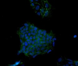
- Immunocytochemistry of fixed and permeabilized 2102EP cells using 10 µg/mL Anti-Human Desmoglein 2 Alexa Fluor® 488. Nuclei are stained with DAPI.
- Conjugate
- Green dye

Supportive validation
- Submitted by
- Invitrogen Antibodies (provider)
- Main image
- Experimental details
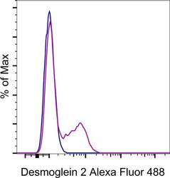
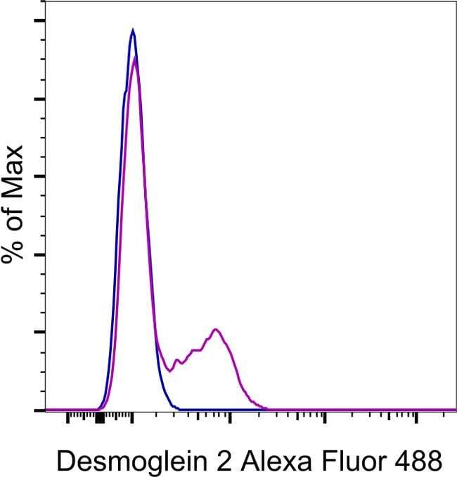
- Staining of a mixture of iPSC and the C2C12 cell line with 0.125 µg of Mouse IgG2b K Isotype Control Alexa Fluor® 488 (Product # 53-4732-80) (blue histogram) or 0.125 µg of Anti-Human Desmoglein 2 Alexa Fluor® 488 (purple histogram). Total viable cells were used for analysis.
- Conjugate
- Green dye
Supportive validation
- Submitted by
- Invitrogen Antibodies (provider)
- Main image
- Experimental details
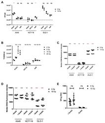
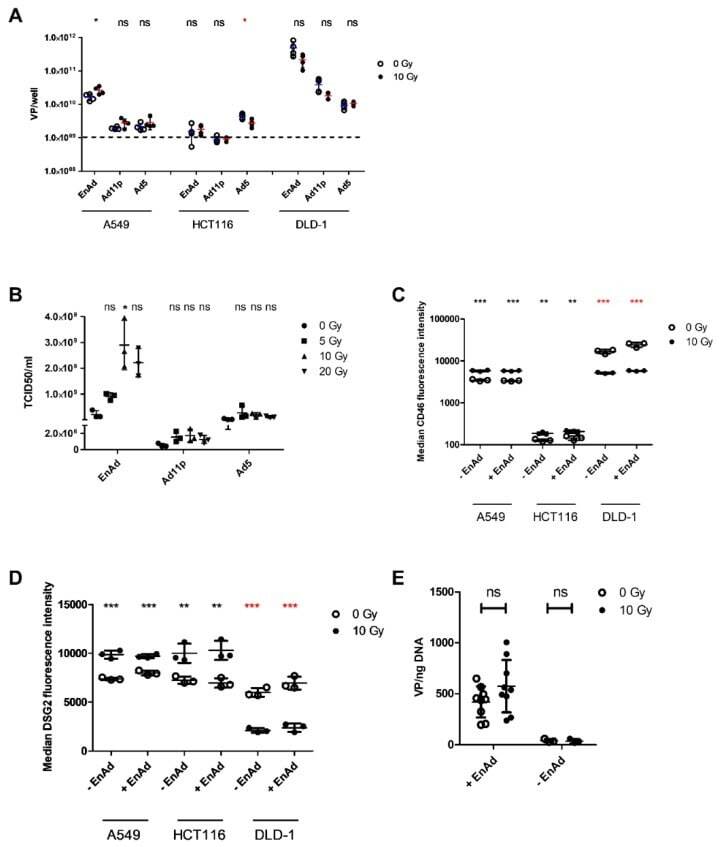
- Figure 1 Irradiation increases EnAd genome replication and particle production. ( A ) Viral genomes were quantified at 72 h post-infection (p. i.) in combined supernatant and lysate of A549, HCT116, and DLD-1 cells mock-infected or infected with EnAd-SA-GFP, Ad11p, or Ad5 at an MOI of 0.1. Cells were being irradiated at 10 Gy 24 h post-infection. Genomes were detected using hexon- (EnAd/Ad11p) or fibre-specific (Ad5) primers and probe. Data represents biological replicates +- SD. ( B ) TCID 50 /mL was quantified for supernatant harvested 5 days p. i. from A549 cells mock-infected or infected with EnAd-SA-GFP, Ad11p or Ad5 and irradiated 24 h p. i. at 0, 5, 10 or 20 Gy. Data shows mean +- SD of three experimental repeats, each point representing the mean of three biological replicates within a single experimental repeat. ( C ) CD46 or ( D ) DSG2 median fluorescence levels were measured 24 h after irradiation. A549, HCT116, or DLD-1 cells were infected with EnAd-SA-GFP at an MOI of 0.1 at 24 h before irradiation with 10 Gy. Data represents biological replicates +- SD of a single representative experiment repeated three times. ( E ) A549 cells were irradiated with 10 Gy 18 h prior to seeding in 12-well plates. At 6 h post-seeding, cells were infected with EnAd-SA-GFP at an MOI of 0 or 0.1. At 2 h post-infection, cells were washed twice before extracting and quantifying DNA for viral genomes by qPCR using hexon-specific primers and probes. Data represent biological replicates +-
- Conjugate
- Green dye
- Submitted by
- Invitrogen Antibodies (provider)
- Main image

- Experimental details
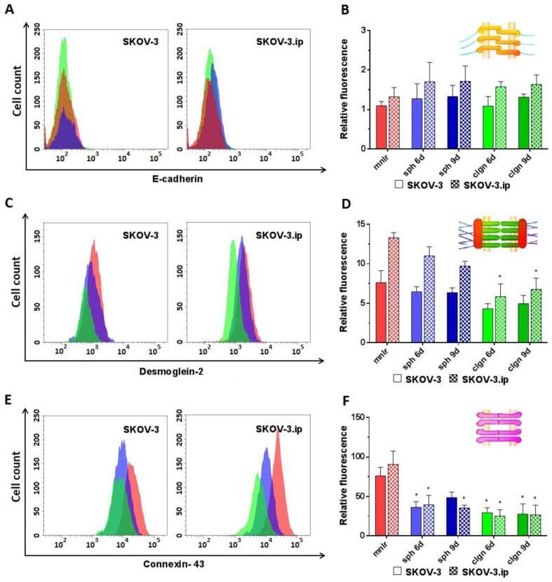
- Figure 3 Expression level of analyzed proteins of adherens junctions (E-cadherin), desmosomes (desmoglein-2) and gap junctions (connexin-43) in SKOV-3 and SKOV-3.ip cells cultured in monolayer and 3D in vitro models. ( A , C , E ) The distributions of SKOV-3 cells (left plot) and SKOV-3.ip cells (right plot) according to fluorescence intensity detected after staining with E-cadherin-specific, desmoglein-2-specific and connexin-43-specific antibodies (red-monolayer culture, blue-spheroids, green-collagen hydrogel); ( B , D , F ) Levels of E-cadherin, desmoglein-2 and connexin-43 in monolayer and 3D models denoted as relative fluorescence values, calculated as a ratio of mean fluorescence intensity of cells stained with specific antibodies to mean fluorescence intensity of cells stained with antibodies of isotypic control. mnlr , monolayer; sph , spheroids; clgn , collagen hydrogel. ""*"" indicates significant difference in RF level from monolayer culture (ANOVA, Holm-Sidak''s multiple comparisons test, p < 0.05).
- Conjugate
- Green dye
- Submitted by
- Invitrogen Antibodies (provider)
- Main image
- Experimental details
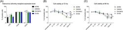
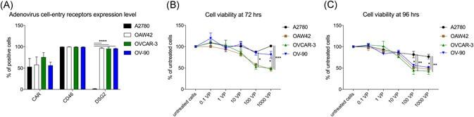
- Figure 1 EOC receptor expression and sensitivity to oncolytic activity to ONCOS-102 treatment. A, Flow cytometry analyses of CAR, CD46, and DSG2 receptor expression on ovarian cancer cells. At least 10 4 events were analyzed for each marker and cell line. Results represent the mean +- SEM of at least two independent experiments. Cell viability at (B) 72 hours or (C) 96 hours after ONCOS-102 treatment in five different concentrations was assessed with the MTS assay. Results are expressed as the mean percent of untreated cells +- SEM. Data represents a pool of two independent experiments run in triplicate. CAR, coxsackie and adenovirus receptor; DSG2, desmoglein-2; EOC, epithelial ovarian cancer
- Conjugate
- Green dye
- Submitted by
- Invitrogen Antibodies (provider)
- Main image

- Experimental details
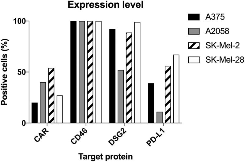
- 10.1080/2162402X.2018.1532763-F0001 Figure 1. Expression of CAR, CD46, Desmoglein-2 and PD-L1 in human melanoma cells measured by flow cytometry (at least 10 4 cells/events were analyzed by flow cytometry in one replicate experiment). Data are expressed as percentage of cells positive for the marker.
- Conjugate
- Green dye