Explore
 Validate
 Learn
Learn Western blot
Antibody data
- Antibody Data
- Antigen structure
- References [0]
- Comments [0]
- Validations
- Western blot [3]
- Immunohistochemistry [4]
Submit
Validation data
Reference
Comment
Report error
- Product number
- NBP1-93704 - Provider product page

- Provider
- Novus Biologicals
- Proper citation
- Novus Cat#NBP1-93704, RRID:AB_11034215
- Product name
- Rabbit Polyclonal MYO1G Antibody
- Antibody type
- Polyclonal
- Description
- Immunogen affinity purified. Specificity of human MYO1G antibody verified on a Protein Array containing target protein plus 383 other non-specific proteins.
- Reactivity
- Human
- Host
- Rabbit
- Isotype
- IgG
- Vial size
- 0.1 ml
- Storage
- Store at 4C short term. Aliquot and store at -20C long term. Avoid freeze-thaw cycles.
No comments: Submit comment
Supportive validation
- Submitted by
- Novus Biologicals (provider)
- Main image

- Experimental details
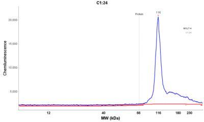
- Simple Western: MYO1G Antibody [NBP1-93704] - Simple Western lane view shows a specific band for MYO1G in 0.2 mg/ml of MOLT-4 lysate(s). This experiment was performed under reducing conditions using the 12-230 kDa separation system.

- Submitted by
- Novus Biologicals (provider)
- Main image
- Experimental details
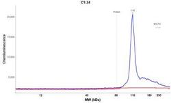
- Simple Western: MYO1G Antibody [NBP1-93704] - Electropherogram image of the corresponding Simple Western lane view. MYO1G antibody was used at 1:25 dilution on MOLT-4 lysate(s) respectively.
- Submitted by
- Novus Biologicals (provider)
- Main image

- Experimental details
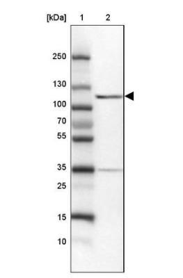
- Western Blot: MYO1G Antibody [NBP1-93704] - Lane 1: Marker [kDa] 250, 130, 100, 70, 55, 35, 25, 15, 10 Lane 2: Human cell line MOLT-4
Supportive validation
- Submitted by
- Novus Biologicals (provider)
- Main image

- Experimental details
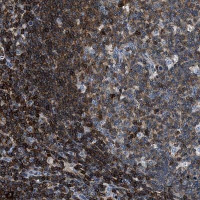
- Immunohistochemistry-Paraffin: MYO1G Antibody [NBP1-93704] - Staining of human lymph node shows strong cytoplasmic positivity in lymphoid cells outside reaction centra.
- Submitted by
- Novus Biologicals (provider)
- Main image

- Experimental details
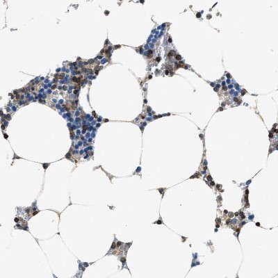
- Immunohistochemistry-Paraffin: MYO1G Antibody [NBP1-93704] - Staining of human bone marrow shows high expression.
- Submitted by
- Novus Biologicals (provider)
- Main image
- Experimental details
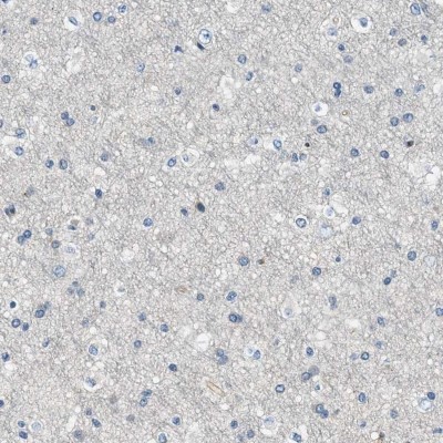
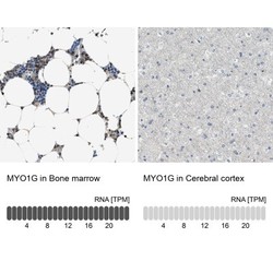
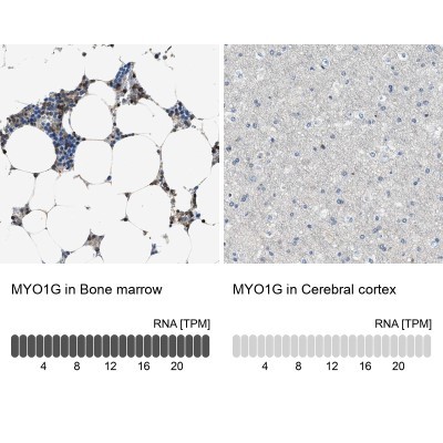
- Immunohistochemistry-Paraffin: MYO1G Antibody [NBP1-93704] - Staining in human bone marrow and cerebral cortex tissues using anti-MYO1G antibody. Corresponding MYO1G RNA-seq data are presented for the same tissues.
- Submitted by
- Novus Biologicals (provider)
- Main image
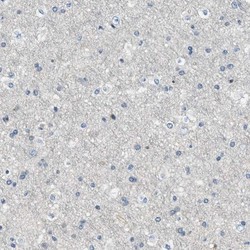
- Experimental details
- Immunohistochemistry-Paraffin: MYO1G Antibody [NBP1-93704] - Staining of human cerebral cortex shows low expression as expected.