 Explore
 Validate
 Learn
Learn Western blot
 ELISA
ELISA Immunocytochemistry
 Immunohistochemistry
Antibody data
- Antibody Data
- Antigen structure
- References [0]
- Comments [0]
- Validations
- Western blot [3]
- Immunohistochemistry [1]
Submit
Validation data
Reference
Comment
Report error
- Product number
- LS-C153997 - Provider product page

- Provider
- LSBio
- Product name
- AKT1 Antibody (phospho-Ser473, clone 17F6.B11) LS-C153997
- Antibody type
- Monoclonal
- Description
- Protein A affinity chromatography
- Reactivity
- Human, Mouse, Rat, Simian
- Host
- Mouse
- Isotype
- IgG
- Antibody clone number
- 17F6.B11
- Storage
- Short term: store at 4°C. Long term: aliquot and store at -20°C. Avoid freeze-thaw cycles.
No comments: Submit comment
Enhanced validation
- Submitted by
- LSBio (provider)
- Enhanced method
- Genetic validation
- Main image

- Experimental details
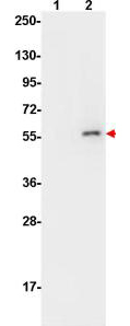
- AKT pS473 Monoclonal Antibody - Western Blot. Anti-AKT pS473 by western blot shows detection of phosphorylated AKT (indicated by arrowhead at ~56 kD) on PDGF stimulated NIH/3T3 cell lysates (lane 2). No reactivity is seen for non-phosphorylated AKT in untreated cells (lane 1). Each lane contained approximately 10 ug of lysate. All samples were loaded on to a 4-20% gradient gel for separation. After electrophoresis, the gel was blocked with 5% BLOTTO (B501-0500 in TBS for 90 min at RT. The membrane was probed with the primary antibody at a 1:10000 dilution in TBS with 0.05% Tween-20 with 1% BSA, for 1 h at 4° C. For detection HRP conjugated Gt-a-Mouse IgG (p/n LS-C60680) was used at a 1:20000 dilution for 1 h at 4° C with FemtoMax enhanced chemiluminescent reagent (p/n FEMTOMAX-100). Images were captured using 2X2 binning for 10-20 sec using a BioSpectrum Imaging System (UVP Ltd.).
- Submitted by
- LSBio (provider)
- Enhanced method
- Genetic validation
- Main image
- Experimental details
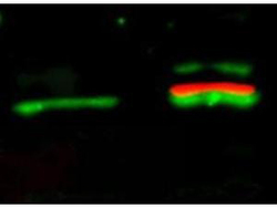
- AKT pS473 Monoclonal Antibody - Western Blot. Anti-AKT pS473 Monoclonal Antibody - Western Blot#Anti-Akt pS473 antibody by fluorescent western blot shows simultaneous detection of unphosphorylated and phosphorylated Akt1 present in serum starved and PDGF stimulated NIH/3T3 whole cell lysates. Lane 1, unstimulated NIH/3T3 lysates contain inactive unphosphorylated Akt1, green band. Lane 2, PDGF stimulated NIH/3T3 lysate contains both inactive (green band) and activated phosphorylated Akt1 (red band). Both lanes were probed with rabbit anti-Akt (pan) and mouse anti-Akt pS473 specific antibodies. This was followed by detection with DyLight 549 conjugated anti-rabbit IgG (green) and DyLight 649 conjugated anti-mouse IgG (red) secondary antibodies.

- Submitted by
- LSBio (provider)
- Enhanced method
- Genetic validation
- Main image
- Experimental details
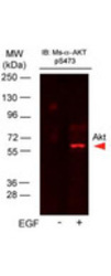
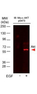
- AKT pS473 Monoclonal Antibody - Western Blot. Anti-AKTpS473 Antibody in western blot showing detection of AKTpS473 in A431 cells. Epidermal growth factor (EGF) was used to stimulate EGF receptor (EGFR) in A431 cells. Cells were stimulated for 15 min with EGF. The Western blot was blocked with Blocking Buffer for Fluorescent Western Blot p/n MB-070, incubated with DyLight649 Conjugated Anti-AKT pS473 Monoclonal Antibody p/n and detected using the BioRad VersaDoc MP4000 system.
Supportive validation
- Submitted by
- LSBio (provider)
- Enhanced method
- Genetic validation
- Main image
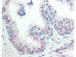
- Experimental details
- AKT pS473 Monoclonal Antibody - Immunohistochemistry. anti-AKT pS473 monoclonal antibody in immunohistochemistry shows detection of phosphorylated AKT pS473 in human prostate tissue. The antibody was used at 20 ug/mL. The staining is much stronger than the weak basal level of phosphorylation in normal prostate tissue. Tissue was formalin fixed and paraffin embedded. No pre-treatment of sample was required.
