Explore
 Validate
 Learn
Learn Western blot
 ELISA
ELISA Immunocytochemistry
Antibody data
- Antibody Data
- Antigen structure
- References [0]
- Comments [0]
- Validations
- Western blot [1]
- Immunohistochemistry [3]
Submit
Validation data
Reference
Comment
Report error
- Product number
- LS-B1183 - Provider product page

- Provider
- LSBio
- Product name
- IHC-plus™ AKT1 Antibody (phospho-Ser473) LS-B1183
- Antibody type
- Polyclonal
- Description
- Immunoaffinity purified
- Reactivity
- Human, Mouse, Rat
- Host
- Rabbit
- Storage
- Store at 4°C or -20°C. Avoid freeze-thaw cycles.
No comments: Submit comment
Supportive validation
- Submitted by
- LSBio (provider)
- Main image

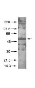
- Experimental details
- Anti-AKT pS473 Antibody - Western Blot. Rabbit anti-AKT pS473 was used at a 1:200 dilution to detect phosphorylated AKT by Western blot. A nuclear extract from cells infected with adenovirus expressing nuclear-targeted AKT kinase was used.
Supportive validation
- Submitted by
- LSBio (provider)
- Main image
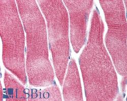
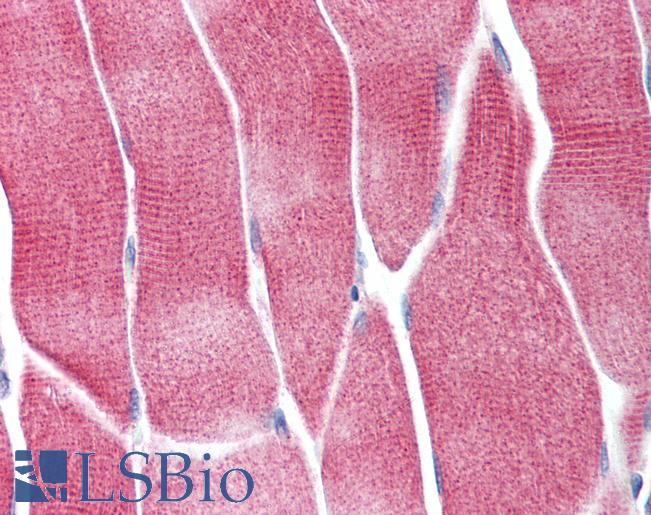
- Experimental details
- Anti-AKT1 antibody IHC of human skeletal muscle. Immunohistochemistry of formalin-fixed, paraffin-embedded tissue after heat-induced antigen retrieval. Antibody concentration 5 ug/ml.
- Submitted by
- LSBio (provider)
- Main image
- Experimental details
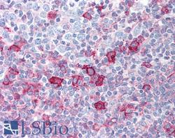
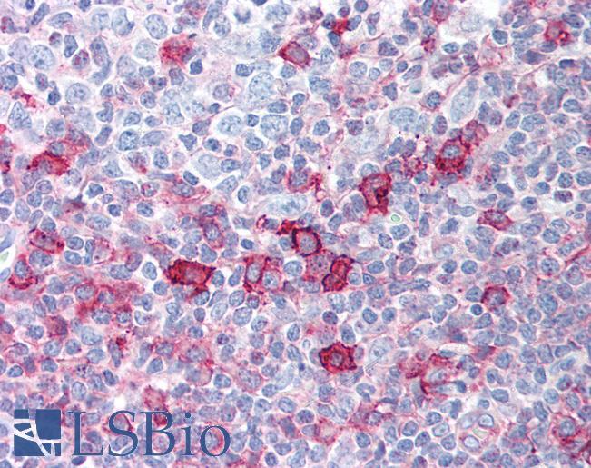
- Anti-AKT1 antibody IHC of human tonsil. Immunohistochemistry of formalin-fixed, paraffin-embedded tissue after heat-induced antigen retrieval. Antibody concentration 5 ug/ml.
- Submitted by
- LSBio (provider)
- Main image
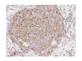
- Experimental details
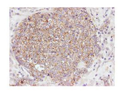
- Immunohistochemistry of Rabbit Anti-Akt pS473 antibody. Tissue: human breast carcinoma. Fixation: formalin fixed paraffin embedded. Antigen retrieval: not required. Primary antibody: Akt pS473 antibody at 100 dilution for 1 h at RT. Secondary antibody: Dako's Techmate streptavidin-biotin reagents at 1:10000 for 45 min at RT. Localization: Akt pS473 is nuclear and occasionally cytoplasmic.