 Explore
 Validate
 Learn
Learn Western blot
 Immunocytochemistry
Antibody data
- Antibody Data
- Antigen structure
- References [0]
- Comments [0]
- Validations
- Western blot [2]
- ELISA [1]
- Immunohistochemistry [3]
Submit
Validation data
Reference
Comment
Report error
- Product number
- NBP1-69924 - Provider product page

- Provider
- Novus Biologicals
- Product name
- Mouse Monoclonal AKT1 Antibody
- Antibody type
- Monoclonal
- Description
- Protein A purified. This AKT phospho Thr308 antibody was purified from concentrated tissue culture supernate by Protein A chromatography. This antibody is specific for human and mouse AKT protein phosphorylated at T308. Cross-reactivity with AKT2 and AKT3 will likely occur.
- Reactivity
- Human, Mouse, Rat, Simian
- Host
- Mouse
- Isotype
- IgG
- Vial size
- 0.1 mg
- Concentration
- 1 mg/ml
- Storage
- Store at -20C. Avoid freeze-thaw cycles.
No comments: Submit comment
Supportive validation
- Submitted by
- Novus Biologicals (provider)
- Main image
- Experimental details
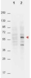
- Western Blot: AKT1 [p Thr308] Antibody (18F3.H11) [NBP1-69924] - Lane 1: non-phosphorylated AKT in untreated cells. Lane 2: phosphorylated AKT on PDGF stimulated NIH/3T3 cell lysates. Load: 15 ug per lane. Primary antibody: AKT phospho Thr308 antibody at a 1:4000 dilution in TBS with 3% BSA, for 3 h at 4 C. Secondary antibody: peroxidase conjugated Gt-a-Mouse IgG (Fc) at a 1:40,000 dilution for 1 h at 4 C. Block: 3% BSA in TBS for 30 min at RT. Predicted/Observed size: (indicated by arrowhead at 56 kDa). Other band(s): unspecific.

- Submitted by
- Novus Biologicals (provider)
- Main image

- Experimental details
- Western Blot: AKT1 [p Thr308] Antibody (18F3.H11) [NBP1-69924] - WB analysis using the Biotin conjugate of AKT1 pT308 antibody. Detection of Lane 1: GST tagged AKT1 active recombinant protein. Lane 2: GST tagged AKT1 inactive recombinant protein. Load: 25 ng per lane. AKT pT308 Biotin conjugated antibody at 1:1000 overnight at 4C. Secondary antibody: HRP Streptavidin.

Supportive validation
- Submitted by
- Novus Biologicals (provider)
- Main image

- Experimental details
- ELISA: AKT1 [p Thr308] Antibody (18F3.H11) [NBP1-69924] - ELISA of AKT1 phospho Thr308 Biotin conjugated antibody. Antigen: Unconjugated AKT1 phospho Thr308 and AKT1 non-phospho Thr308. Coating amount: 0.1 ug per well. Primary antibody: AKT1 phospho Thr308 Biotin conjugated antibody at 5 ug/mL. Dilution series: 3-fold. Mid-point concentration: 5 ng/mL AKT1 phospho Thr308 Biotin conjugated antibody. Secondary antibody: Peroxidase streptavidin secondary antibody at 1:10,000. Substrate: TMB

Supportive validation
- Submitted by
- Novus Biologicals (provider)
- Main image

- Experimental details
- Immunohistochemistry-Paraffin: AKT1 [p Thr308] Antibody (18F3.H11) [NBP1-69924] - Analysis of Biotin conjugate of NBP1-69924. 20 ug/mL for 1 h at RT Secondary antibody: Streptavidin Conj. HRP 10 ug/ml Localization: nuclear and occasionally cytoplasmic Staining: antibody as precipitated red signal with a hematoxylin purple nuclear count

- Submitted by
- Novus Biologicals (provider)
- Main image

- Experimental details
- Immunohistochemistry-Paraffin: AKT1 [p Thr308] Antibody (18F3.H11) [NBP1-69924] - IHC analysis. Detection of phosphorylated AKT phospho Thr308 in human FFPE brain cerebellum tissue (40X). AKT1 phospho Thr308 antibody was used at 20 ug/mL. The image shows strong staining of Purkinje neurons. Red: AKT1 pT308. Purple: Hematoxylin nuclear counterstain.

- Submitted by
- Novus Biologicals (provider)
- Main image

- Experimental details
- Immunohistochemistry-Paraffin: AKT1 [p Thr308] Antibody (18F3.H11) [NBP1-69924] - Analysis using the Biotin conjugate of NBP1-69924. Tissue: Human Skin at pH6. Fixation: formalin fixed paraffin embedded. Primary antibody: Collagen Type I antibody at 10 ug/mL for 1 h at RT. Localization: Collagen Type I is secreted in the extracellular
