Explore
 Validate
 Learn
Learn Western blot
Antibody data
- Antibody Data
- Antigen structure
- References [0]
- Comments [0]
- Validations
- Western blot [1]
- ELISA [1]
- Immunohistochemistry [2]
Submit
Validation data
Reference
Comment
Report error
- Product number
- NBP2-21679 - Provider product page

- Provider
- Novus Biologicals
- Product name
- Mouse Monoclonal AKT1 Antibody
- Antibody type
- Monoclonal
- Description
- Protein A purified. This AKT phospho Thr308 antibody was purified from concentrated tissue culture supernate by Protein A chromatography. This antibody is specific for human and mouse AKT protein phosphorylated at T308. Cross-reactivity with AKT2 and AKT3 will likely occur.
- Reactivity
- Human, Mouse, Rat, Simian
- Host
- Mouse
- Conjugate
- Biotin
- Isotype
- IgG
- Vial size
- 0.05 mg
- Concentration
- LYOPH
- Storage
- Store lyophilized antibody at 4C. Aliquot reconstituted liquid and store at -20C. Avoid freeze-thaw cycles.
No comments: Submit comment
Supportive validation
- Submitted by
- Novus Biologicals (provider)
- Main image
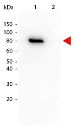
- Experimental details
- Western Blot: AKT1 [p Thr308] Antibody (18F3.H11) [Biotin] [NBP2-21679] - Lane 1: GST-tagged AKT1 active recombinant protein. Lane 2: GST-tagged AKT1 inactive recombinant protein. Load: 25 ng per lane. AKT1 phospho Thr308 Biotin conjugated antibody at 1:1000 overnight at 4C. Secondary antibody: HRP Streptavidin secondary antibody at 1:40,000 for 30 min at RT. Block: MB-070 for 30 min at RT. Predicted/Observed size: 79 kDa, 79 kDa for AKT phospho Thr308.

Supportive validation
- Submitted by
- Novus Biologicals (provider)
- Main image

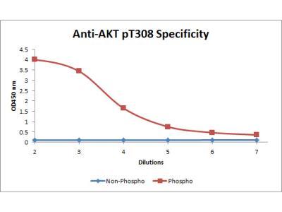
- Experimental details
- ELISA: AKT1 [p Thr308] Antibody (18F3.H11) [Biotin] [NBP2-21679] - ELISA of AKT1 phospho Thr308 Biotin conjugated antibody. Antigen: Unconjugated AKT1 phospho Thr308 and AKT1 non-phospho Thr308. Coating amount: 0.1 ug per well. Primary antibody: AKT1 phospho Thr308 Biotin conjugated antibody at 5 ug/mL. Dilution series: 3-fold. Mid-point concentration: 5 ng/mL AKT1 phospho Thr308 Biotin conjugated antibody. Secondary antibody: Peroxidase streptavidin secondary antibody at 1:10,000. Substrate: TMB
Supportive validation
- Submitted by
- Novus Biologicals (provider)
- Main image

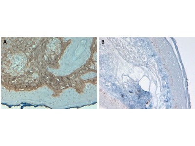
- Experimental details
- Immunohistochemistry: Akt1 [p Thr308] Antibody (18F3.H11) [Biotin] [NBP2-21679] - Tissue: Human Skin at pH9. Fixation: formalin fixed paraffin embedded. Primary antibody: Collagen Type I antibody at 10 ug/mL for 1 h at RT. Localization: Collagen Type I is secreted in the extracellular matrix. Staining: Collagen Type I as precipitated brown signal (A) with hematoxylin purple nuclear counterstain. With corresponding negative conrol (B).
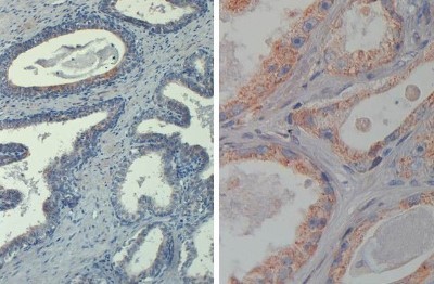
- Submitted by
- Novus Biologicals (provider)
- Main image
- Experimental details
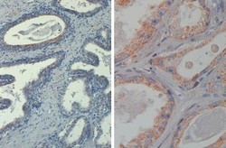
- Immunohistochemistry: AKT1 [p Thr308] Antibody (18F3.H11) [Biotin] [NBP2-21679] - IHC-P analysis of FFPE human prostate tissue using AKT1 phospho Thr308 antibody at 20 ug/mL. Left panel: 20X magnification. Right panel: 40X magnification. Heat antigen retrieval in citrate buffer pH 6.2. Secondary antibody: streptavidin-HRP at 10 ug/mL. Antibody as precipitated red signal with hemotoxylin purple nuclear counterstain.