Explore
 Validate
 Learn
Learn Western blot
Antibody data
- Antibody Data
- Antigen structure
- References [0]
- Comments [0]
- Validations
- Western blot [2]
- Immunocytochemistry [1]
- Immunohistochemistry [1]
- Flow cytometry [1]
Submit
Validation data
Reference
Comment
Report error
- Product number
- AP7141a - Provider product page

- Provider
- Abcepta
- Proper citation
- Abgent Cat#AP7141a, RRID:AB_889332
- Product name
- AKT1 Antibody (N-term)
- Antibody type
- Polyclonal
- Antigen
- Synthetic peptide
- Description
- Purified Rabbit Polyclonal Antibody (Pab)
- Reactivity
- Human
- Host
- Rabbit
- Conjugate
- Unconjugated
- Isotype
- IgG
- Antibody clone number
- RB11644
- Vial size
- 400 µl
- Storage
- Store at 2 to 8°C.Antibody is stable for 49 months.
No comments: Submit comment
Supportive validation
- Submitted by
- Abcepta (provider)
- Main image
- Experimental details
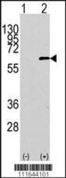
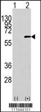
- Western blot analysis of AKT1 Antibody (N-term) polyclonal antibody(Cat.#AP7141a)(arrow). 293 cell lysates (2 ug/lane) either nontransfected (Lane 1) or transiently transfected with the AKT1 gene (Lane 2) (Origene Technologies).
- Primary Ab dilution
- 1:1000
- Submitted by
- Abcepta (provider)
- Main image
- Experimental details
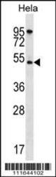
- AKT1 Antibody (N-term) (Cat. #AP7141a) western blot analysis in Hela cell line lysates (35ug/lane).This demonstrates the AKT1 antibody detected the AKT1 protein (arrow).
- Primary Ab dilution
- 1:1000

Supportive validation
- Submitted by
- Abcepta (provider)
- Main image
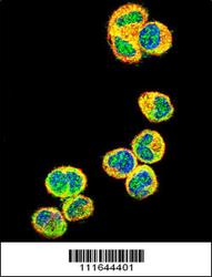
- Experimental details
- Confocal immunofluorescent analysis of AKT1 Antibody (N-term)(Cat#AP7141a) with MDA-MB435 cell followed by Alexa Fluor 488-conjugated goat anti-rabbit lgG (green).Actin filaments have been labeled with Alexa Fluor 555 phalloidin (red).DAPI was used to stain the cell nuclear (blue).
- Primary Ab dilution
- 1:10~50

Supportive validation
- Submitted by
- Abcepta (provider)
- Main image
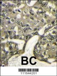
- Experimental details
- "Formalin-fixed and paraffin-embedded human breast carcinoma reacted with AKT1 antibody (N-term)(Cat.#AP7141a), which was peroxidase-conjugated to the secondary antibody, followed by DAB staining. This data demonstrates the use of this antibody for immunohistochemistry; clinical relevance has not been evaluated."
- Primary Ab dilution
- 1:50~100

Supportive validation
- Submitted by
- Abcepta (provider)
- Main image

- Experimental details
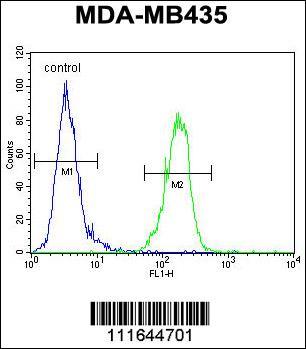
- AKT1 Antibody (N-term) (Cat. #AP7141a) flow cytometric analysis of MDA-MB435 cells (right histogram) compared to a negative control cell (left histogram).FITC-conjugated goat-anti-rabbit secondary antibodies were used for the analysis.
- Primary Ab dilution
- 1:10~50