Explore
 Validate
 Learn
Learn Western blot
 ELISA
ELISAAntibody data
- Antibody Data
- Antigen structure
- References [2]
- Comments [0]
- Validations
- Western blot [2]
- Immunocytochemistry [1]
- Immunohistochemistry [1]
Submit
Validation data
Reference
Comment
Report error
- Product number
- GTX28805 - Provider product page

- Provider
- GeneTex
- Proper citation
- GeneTex Cat#GTX28805, RRID:AB_367524
- Product name
- AKT antibody
- Antibody type
- Polyclonal
- Antigen
- Peptide (available as GTX29041) C-terminus (460-480) of human, rat and mouse and chicken Akt proteins. This whole rabbit serum was prepared by repeated immunizations with a KLH conjugated peptide.
- Host
- Rabbit
- Isotype
- IgG
- Vial size
- 100µl
- Concentration
- Whole antiserum
- Storage
- Upon Receipt - Keep as concentrated solution. Aliquot and store at -20°C or below. Avoid freeze-thaw cycles.
Submitted references Orchiectomy and letrozole differentially regulate synaptic plasticity and spatial memory in a manner that is mediated by SRC-1 in the hippocampus of male mice.
PFKFB3 modulates glycolytic metabolism and alleviates endoplasmic reticulum stress in human osteoarthritis cartilage.
Zhao J, Bian C, Liu M, Zhao Y, Sun T, Xing F, Zhang J
The Journal of steroid biochemistry and molecular biology 2018 Apr;178:354-368
The Journal of steroid biochemistry and molecular biology 2018 Apr;178:354-368
PFKFB3 modulates glycolytic metabolism and alleviates endoplasmic reticulum stress in human osteoarthritis cartilage.
Qu J, Lu D, Guo H, Miao W, Wu G, Zhou M
Clinical and experimental pharmacology & physiology 2016 Mar;43(3):312-8
Clinical and experimental pharmacology & physiology 2016 Mar;43(3):312-8
No comments: Submit comment
Supportive validation
- Submitted by
- GeneTex (provider)
- Main image
- Experimental details
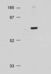
- Western blot using Akt antibody at 1/500 on 20 ug NIH/3T3 whole cell lysate
- Validation comment
- WB

- Submitted by
- GeneTex (provider)
- Main image
- Experimental details
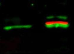
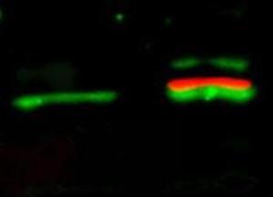
- Western Blot of simultaneous detection of unphosphorylated and phosphorylated Rabbit Anti-AKT antibody (GTX28805). Lane 1: unstimulated NIH/3T3 lysates contain inactive unphosphorylated Akt1, green band. Lane 2: PDGF stimulated NIH/3T3 lysate contains both inactive (green band) and activated phosphorylated Akt1 (red band). Load: 35 µg per lane. Primary antibody: rabbit anti-Akt (pan) and mouse anti-Akt pS473 specific antibodies at 1:1000 for overnight at 4°C. Secondary antibody: Flourescent 549 conjugated anti-rabbit IgG (green) and flourescent 649 conjugated anti-mouse IgG (red) secondary antibodies at 1:10,000 for 45 min at RT. Block: 5% BLOTTO overnight at 4°C.
- Validation comment
- WB
Supportive validation
- Submitted by
- GeneTex (provider)
- Main image
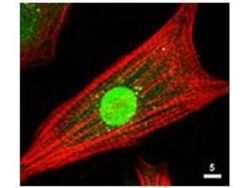
- Experimental details
- ICC/IF of the Akt antibody is used at 1: 80 for 1hr at RT on cultured neonatal rat cardiomyocytes (Green). The red is Texas-red phalloidin that labels actin filaments.

Supportive validation
- Submitted by
- GeneTex (provider)
- Main image
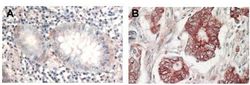
- Experimental details
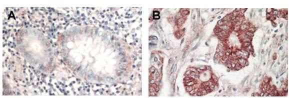
- Immunohistochemical analysis of formalin-fixed paraffin-embedded tissue, A: Normal colon tissue, B: Colon tumor tissue, using AKT(GTX28805) antibody at 1:1,000 dilution for 1 h at RT. Peroxidase rabbit secondary antibody at 1:10,000 for 45 min at RT. Localization: AKT is nuclear. Staining: AKT as precipitated red signal with hematoxylin purple nuclear counterstain.