Explore
 Validate
 Learn
Learn Western blot
 ELISA
ELISA Immunocytochemistry
Antibody data
- Antibody Data
- Antigen structure
- References [14]
- Comments [0]
- Validations
- Immunocytochemistry [4]
- Immunohistochemistry [1]
- Flow cytometry [4]
- Other assay [5]
Submit
Validation data
Reference
Comment
Report error
- Product number
- 700392 - Provider product page

- Provider
- Invitrogen Antibodies
- Product name
- Phospho-AKT1 (Ser473) Recombinant Rabbit Monoclonal Antibody (98H9L8)
- Antibody type
- Monoclonal
- Antigen
- Synthetic peptide
- Description
- This antibody is predicted to react with bovine, canine, chicken, chimpanzee, equine, feline, rat, Rhesus monkey , Xenopus and zebrafish based on sequence homology. Intact IgG appears on a non-reducing gel as ~150 kDa band and upon reduction generating a ~25 kDa light chain band and a ~50 kDa heavy chain. Recombinant rabbit monoclonal antibodies are produced using in vitro expression systems. The expression systems are developed by cloning in the specific antibody DNA sequences from immunoreactive rabbits. Then, individual clones are screened to select the best candidates for production. The advantages of using recombinant rabbit monoclonal antibodies include: better specificity and sensitivity, lot-to-lot consistency, animal origin-free formulations, and broader immunoreactivity to diverse targets due to larger rabbit immune repertoire.
- Reactivity
- Human, Mouse
- Host
- Rabbit
- Isotype
- IgG
- Antibody clone number
- 98H9L8
- Vial size
- 100 μg
- Concentration
- 0.5 mg/mL
- Storage
- Store at 4°C short term. For long term storage, store at -20°C, avoiding freeze/thaw cycles.
Submitted references Empagliflozin maintains capillarization and improves cardiac function in a murine model of left ventricular pressure overload.
Exhausted CD4(+) T Cells during Malaria Exhibit Reduced mTORc1 Activity Correlated with Loss of T-bet Expression.
AKT signaling promotes DNA damage accumulation and proliferation in polycystic kidney disease.
Preconception paternal alcohol exposure exerts sex-specific effects on offspring growth and long-term metabolic programming.
Programmed increases in LXRα induced by paternal alcohol use enhance offspring metabolic adaptation to high-fat diet induced obesity.
Significance of PYK2 level as a prognosis predictor in patients with colon adenocarcinoma after surgical resection.
Naive CD4(+) T Cells Carrying a TLR2 Agonist Overcome TGF-β-Mediated Tumor Immune Evasion.
Synergistic Activity of Deguelin and Fludarabine in Cells from Chronic Lymphocytic Leukemia Patients and in the New Zealand Black Murine Model.
Snail heterogeneity in clear cell renal cell carcinoma.
Harnessing Connectivity in a Large-Scale Small-Molecule Sensitivity Dataset.
Intramembrane binding of VE-cadherin to VEGFR2 and VEGFR3 assembles the endothelial mechanosensory complex.
Expression of EGFR and molecules downstream to PI3K/Akt, Raf-1-MEK-1-MAP (Erk1/2), and JAK (STAT3) pathways in invasive lung adenocarcinomas resected at a single institution.
Experimentally validated novel inhibitors of Helicobacter pylori phosphopantetheine adenylyltransferase discovered by virtual high-throughput screening.
Nanoparticle-mediated signaling endosome localization regulates growth cone motility and neurite growth.
Nakao M, Shimizu I, Katsuumi G, Yoshida Y, Suda M, Hayashi Y, Ikegami R, Hsiao YT, Okuda S, Soga T, Minamino T
Scientific reports 2021 Sep 15;11(1):18384
Scientific reports 2021 Sep 15;11(1):18384
Exhausted CD4(+) T Cells during Malaria Exhibit Reduced mTORc1 Activity Correlated with Loss of T-bet Expression.
Villegas-Mendez A, Khandelwal G, McGowan LM, Dookie RS, Haley MJ, George C, Sims D, Lord GM, Sinclair LV, Jenner RG, Couper KN
Journal of immunology (Baltimore, Md. : 1950) 2020 Sep 15;205(6):1608-1619
Journal of immunology (Baltimore, Md. : 1950) 2020 Sep 15;205(6):1608-1619
AKT signaling promotes DNA damage accumulation and proliferation in polycystic kidney disease.
Conduit SE, Davies EM, Ooms LM, Gurung R, McGrath MJ, Hakim S, Cottle DL, Smyth IM, Dyson JM, Mitchell CA
Human molecular genetics 2020 Jan 1;29(1):31-48
Human molecular genetics 2020 Jan 1;29(1):31-48
Preconception paternal alcohol exposure exerts sex-specific effects on offspring growth and long-term metabolic programming.
Chang RC, Wang H, Bedi Y, Golding MC
Epigenetics & chromatin 2019 Jan 22;12(1):9
Epigenetics & chromatin 2019 Jan 22;12(1):9
Programmed increases in LXRα induced by paternal alcohol use enhance offspring metabolic adaptation to high-fat diet induced obesity.
Chang RC, Thomas KN, Bedi YS, Golding MC
Molecular metabolism 2019 Dec;30:161-172
Molecular metabolism 2019 Dec;30:161-172
Significance of PYK2 level as a prognosis predictor in patients with colon adenocarcinoma after surgical resection.
Liu S, Chen L, Xu Y
OncoTargets and therapy 2018;11:7625-7634
OncoTargets and therapy 2018;11:7625-7634
Naive CD4(+) T Cells Carrying a TLR2 Agonist Overcome TGF-β-Mediated Tumor Immune Evasion.
Ibrahim M, Scozzi D, Toth KA, Ponti D, Kreisel D, Menna C, De Falco E, D'Andrilli A, Rendina EA, Calogero A, Krupnick AS, Gelman AE
Journal of immunology (Baltimore, Md. : 1950) 2018 Jan 15;200(2):847-856
Journal of immunology (Baltimore, Md. : 1950) 2018 Jan 15;200(2):847-856
Synergistic Activity of Deguelin and Fludarabine in Cells from Chronic Lymphocytic Leukemia Patients and in the New Zealand Black Murine Model.
Rebolleda N, Losada-Fernandez I, Perez-Chacon G, Castejon R, Rosado S, Morado M, Vallejo-Cremades MT, Martinez A, Vargas-Nuñez JA, Perez-Aciego P
PloS one 2016;11(4):e0154159
PloS one 2016;11(4):e0154159
Snail heterogeneity in clear cell renal cell carcinoma.
Zaldumbide L, Erramuzpe A, Guarch R, Pulido R, Cortés JM, López JI
BMC cancer 2016 Mar 8;16:194
BMC cancer 2016 Mar 8;16:194
Harnessing Connectivity in a Large-Scale Small-Molecule Sensitivity Dataset.
Seashore-Ludlow B, Rees MG, Cheah JH, Cokol M, Price EV, Coletti ME, Jones V, Bodycombe NE, Soule CK, Gould J, Alexander B, Li A, Montgomery P, Wawer MJ, Kuru N, Kotz JD, Hon CS, Munoz B, Liefeld T, Dančík V, Bittker JA, Palmer M, Bradner JE, Shamji AF, Clemons PA, Schreiber SL
Cancer discovery 2015 Nov;5(11):1210-23
Cancer discovery 2015 Nov;5(11):1210-23
Intramembrane binding of VE-cadherin to VEGFR2 and VEGFR3 assembles the endothelial mechanosensory complex.
Coon BG, Baeyens N, Han J, Budatha M, Ross TD, Fang JS, Yun S, Thomas JL, Schwartz MA
The Journal of cell biology 2015 Mar 30;208(7):975-86
The Journal of cell biology 2015 Mar 30;208(7):975-86
Expression of EGFR and molecules downstream to PI3K/Akt, Raf-1-MEK-1-MAP (Erk1/2), and JAK (STAT3) pathways in invasive lung adenocarcinomas resected at a single institution.
Torres AF, Nogueira C, Magalhaes J, Costa IS, Aragao A, Gomes Neto A, Martins F, Tavora F
Analytical cellular pathology (Amsterdam) 2014;2014:352925
Analytical cellular pathology (Amsterdam) 2014;2014:352925
Experimentally validated novel inhibitors of Helicobacter pylori phosphopantetheine adenylyltransferase discovered by virtual high-throughput screening.
Cheng CS, Jia KF, Chen T, Chang SY, Lin MS, Yin HS
PloS one 2013;8(9):e74271
PloS one 2013;8(9):e74271
Nanoparticle-mediated signaling endosome localization regulates growth cone motility and neurite growth.
Steketee MB, Moysidis SN, Jin XL, Weinstein JE, Pita-Thomas W, Raju HB, Iqbal S, Goldberg JL
Proceedings of the National Academy of Sciences of the United States of America 2011 Nov 22;108(47):19042-7
Proceedings of the National Academy of Sciences of the United States of America 2011 Nov 22;108(47):19042-7
No comments: Submit comment
Supportive validation
- Submitted by
- Invitrogen Antibodies (provider)
- Main image

- Experimental details
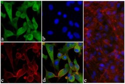
- Immunofluorescent analysis of Phospho-AKT pSer473 in mouse fibroblasts cells treated with 10 µg/mL Insulin (top right) or untreated (top left) using a Phospho-AKT pSer473 recombinant rabbit monoclonal antibody (Product # 700392) at a dilution of 5 µg/mL followed by detection using an Alexa Fluor 488-conjugated goat anti-rabbit secondary antibody at a dilution of 1:1000 and nuclei staining using Hoechst (blue). Signal is knocked down after incubation with the phosphopeptide used as an immunogen (bottom left) but not with the non-phosphopeptide (bottom right).

- Submitted by
- Invitrogen Antibodies (provider)
- Main image
- Experimental details
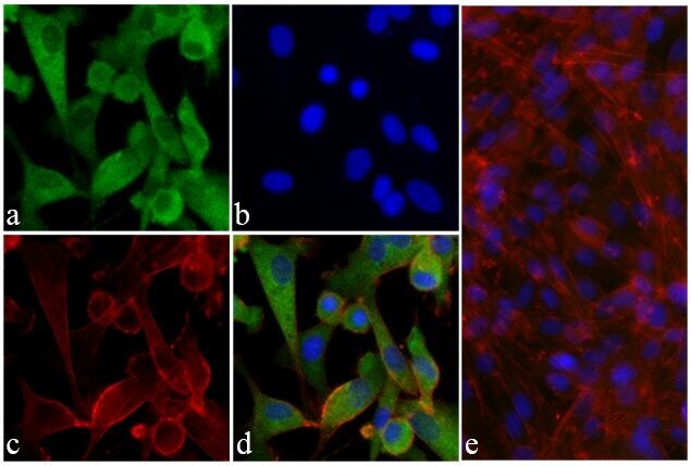
- Immunofluorescent analysis of AKT (pS473) was done on 70% confluent log phase U-87 MG cells. The cells were fixed with 4% paraformaldehyde for 15 minutes; permeabilized with 0.25% Triton X-100 for 10 minutes followed by blocking with 5% BSA for 1 hour at room temperature. The cells were incubated with AKT (pS473) Recombinant Rabbit Monoclonal Antibody (Product # 700392) at 1 µg-2 µg in 1% BSA and incubated for 3 hours at room temperature and then labeled with Alexa Fluor® 488 Goat anti-Rabbit IgG Secondary Antibody (Product # A-11008) at a dilution of 1:400 for 30 minutes at room temperature (Panel a: green). Nuclei (Panel b: blue) were stained with SlowFade® Gold Antifade Mountant with DAPI (Product # S36938). F-actin (Panel c: red) was stained with Alexa Fluor® 594 Phalloidin (Product # A12381). Panel d is a merged image showing cytoplasmic localization of AKT (pS473). Panel e shows competition with AKT (pS473) peptide. The images were captured at 20X magnification.

- Submitted by
- Invitrogen Antibodies (provider)
- Main image

- Experimental details
- Immunofluorescent analysis of AKT (pS473) was done on 70% confluent log phase U-87 MG cells. The cells were fixed with 4% paraformaldehyde for 15 minutes; permeabilized with 0.25% Triton X-100 for 10 minutes followed by blocking with 5% BSA for 1 hour at room temperature. The cells were incubated with AKT (pS473) Recombinant Rabbit Monoclonal Antibody (Product # 700392) at 1 µg-2 µg in 1% BSA and incubated for 3 hours at room temperature and then labeled with Alexa Fluor® 488 Goat anti-Rabbit IgG Secondary Antibody (Product # A-11008) at a dilution of 1:400 for 30 minutes at room temperature (Panel a: green). Nuclei (Panel b: blue) were stained with SlowFade® Gold Antifade Mountant with DAPI (Product # S36938). F-actin (Panel c: red) was stained with Alexa Fluor® 594 Phalloidin (Product # A12381). Panel d is a merged image showing cytoplasmic localization of AKT (pS473). Panel e shows competition with AKT (pS473) peptide. The images were captured at 20X magnification.
- Submitted by
- Invitrogen Antibodies (provider)
- Main image
- Experimental details
- Immunofluorescent analysis of Phospho-AKT pSer473 in mouse fibroblasts cells treated with 10 µg/mL Insulin (top right) or untreated (top left) using a Phospho-AKT pSer473 recombinant rabbit monoclonal antibody (Product # 700392) at a dilution of 5 µg/mL followed by detection using an Alexa Fluor 488-conjugated goat anti-rabbit secondary antibody at a dilution of 1:1000 and nuclei staining using Hoechst (blue). Signal is knocked down after incubation with the phosphopeptide used as an immunogen (bottom left) but not with the non-phosphopeptide (bottom right).

Supportive validation
- Submitted by
- Invitrogen Antibodies (provider)
- Main image

- Experimental details
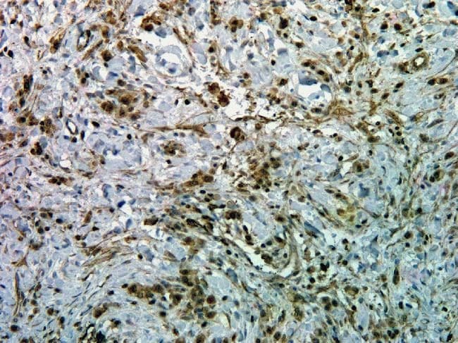
- Immunohistochemistry analysis of Phospho-AKT pSer473 in paraffin-embedded human esophagus carninoma using a Phospho-AKT pSer473 monoclonal antibody (Product # 700392) at a dilution of 0.5 µg/mL. Tissues were pretreated with EDTA and staining was visualized using DAB. Images were taken at a magnification of 20x. Results show nuclear and cytoplasmic staining in tumor cells.
Supportive validation
- Submitted by
- Invitrogen Antibodies (provider)
- Main image
- Experimental details
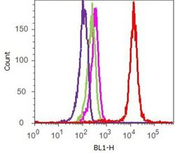
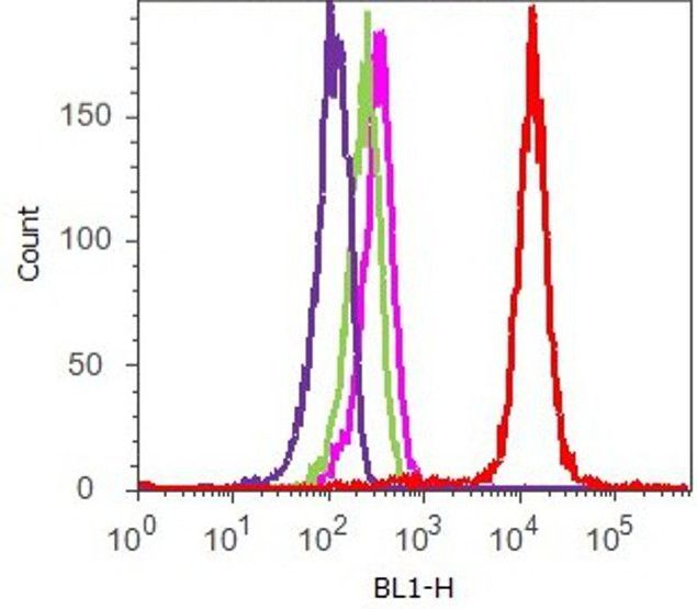
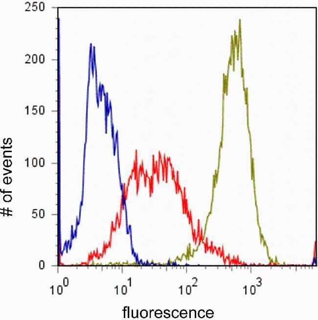
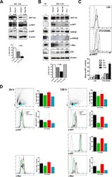
- Flow cytometry analysis of AKT [pS473] was done on U-87 MG cells. Cells were fixed with 70% ethanol for 10 minutes, permeabilized with 0.25% Tritonª X-100 for 20 minutes, and blocked with 5% BSA for 1 hour at room temperature. Cells were labeled with ABfinityª AKT [pS473] Recombinant Rabbit Monoclonal Antibody (700392, red histogram) or with rabbit isotype control (pink histogram) at 2.5 µg/million cells in 2.5% BSA. After incubation at room temperature for 2-3 hours, the cells were labeled with Alexa Fluor¨ 488 Goat Anti-Rabbit Secondary Antibody (A11008) at a dilution of 1:400 for 30 minutes at room temperature. The representative 10,000 cells were acquired and analyzed for each sample using an Attune¨ Acoustic Focusing Cytometer. The purple histogram represents unstained control cells and the green histogram represents no-primary-antibody control.
- Submitted by
- Invitrogen Antibodies (provider)
- Main image
- Experimental details
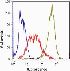
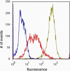
- Flow cytometry analysis of Phospho-AKT pSer473 in Jurkat cells incubated with 50uM LY294002 for 1hr (red) or untreated (green) using a Phospho-AKT pSer473 recombinant rabbit monoclonal antibody (Product # 700392) at a dilution of 1 µg. Cells were fixed and permeabilized using FIX & PERM (Product # GAS-004) reagent, and detection was performed using an Alexa Fluor 488 goat anti-rabbit IgG compared to a control without primary antibody (blue).
- Submitted by
- Invitrogen Antibodies (provider)
- Main image
- Experimental details
- Flow cytometry analysis of Phospho-AKT pSer473 in Jurkat cells incubated with 50uM LY294002 for 1hr (red) or untreated (green) using a Phospho-AKT pSer473 recombinant rabbit monoclonal antibody (Product # 700392) at a dilution of 1 µg. Cells were fixed and permeabilized using FIX & PERM (Product # GAS-004) reagent, and detection was performed using an Alexa Fluor 488 goat anti-rabbit IgG compared to a control without primary antibody (blue).

- Submitted by
- Invitrogen Antibodies (provider)
- Main image

- Experimental details
- Flow cytometry analysis of Phospho-AKT pSer473 in Jurkat cells incubated with 50uM LY294002 for 1hr (red) or untreated (green) using a Phospho-AKT pSer473 recombinant rabbit monoclonal antibody (Product # 700392) at a dilution of 1 µg. Cells were fixed and permeabilized using FIX & PERM (Product # GAS-004) reagent, and detection was performed using an Alexa Fluor 488 goat anti-rabbit IgG compared to a control without primary antibody (blue).

Supportive validation
- Submitted by
- Invitrogen Antibodies (provider)
- Main image

- Experimental details
- NULL
- Submitted by
- Invitrogen Antibodies (provider)
- Main image
- Experimental details
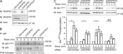
- Figure 5. VEGFRs in PI 3-kinase and integrin activation. (A) VEGFR knockdown. HUVECs transfected with the indicated siRNAs for 72 h were analyzed for VEGFR expression by immunoblotting. Three independent experiments gave similar results. (B) PI3K signaling. HUVECs transfected as in A were subjected to shear stress and p85 was immunoprecipitated. Samples were then analyzed by immunoblotting and densitometry as in Fig 2 C . Values are means +- SEM, n = 4. (C) Akt signaling. siRNA-treated HUVECs were subjected to laminar shear stress for the indicated times and lysates were analyzed by immunoblotting with the indicated antibodies to determine Akt activity. Results were quantified by densitometry, with actin serving as a loading control. Values are means +- SEM (error bars), n = 3. *, P < 0.05 relative to unstimulated cells by two-way ANOVA. IB, immunoblotting.

- Submitted by
- Invitrogen Antibodies (provider)
- Main image
- Experimental details
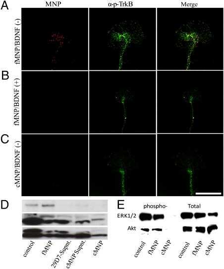
- Fig 4 Deguelin downregulates AKT/NFkappaB mediated prosurvival signals in CLL cells. (A) PBMCs from 3 CLL patients were treated for 24h with 0, 10, or 100 muM deguelin. Viability was evaluated by flow cytometry after Annexin V/IP staining. Figure shows immunoreactive bands for several deguelin target molecules and beta-actin in western blots from a representative sample. Numbers indicate the signal intensity of beta-actin-normalized bands from drug treated samples compared to the untreated ones (Control). (B) PBMCs from two CLL patients were treated with 0, 10, or 100 muM deguelin or 1 mug/ml fludarabine and co-cultured with Ltk - 24h. Cell viability assays and western blot from a representative sample were done as in (A). (C) PBMCs from 2 CLL patients were co-cultured with Ltk - or 3T3-CD40L cells and p-AKT expression was evaluated in CLL cells by intracellular staining and flow cytometry at different time points. Histograms show the stains in one of the samples (solid lines from left to right: p-AKT staining at 0, 24, 48 and 120h; dotted lines from left to right: unstained control (samples without primary antibody plus secondary antibody) and isotype control. The lower graph shows the differences in the Mean Fluorescence Intensity (MFI, in channel numbers) of p-AKT compared to the unstained control in both samples (bars show mean and SD). The dotted horizontal line indicates the mean difference in MFI (10 +- 5) between the isotype controls and corresponding unstained contro

- Submitted by
- Invitrogen Antibodies (provider)
- Main image

- Experimental details
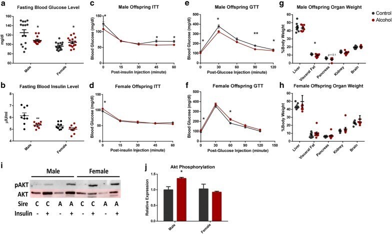
- Fig. 2 Chronic preconception male ethanol exposure exerts sex-specific effects on offspring metabolic function. a Fasting blood glucose levels compared between preconception treatments ( n = 15 males, 15 females). b Fasting insulin levels compared between preconception treatments ( n = nine males, nine females). c - f Glucose tolerance (GTT) and insulin tolerance (ITT) tests in the offspring of ethanol-exposed and control males ( n = 15 males, 15 females). Organ weights of g male and h female offspring compared between the two preconception treatment groups ( n = nine males, nine females). i Representative immunoblot comparing total and phosphorylated AKT (Ser473) between male and female offspring sired by ethanol-exposed and control males ( n = six males and six females). j Densitometry analysis of immunoblots comparing total and phosphorylated AKT ( n = six males and six females). Data analyzed using either an unpaired t test or a two-way ANOVA followed by Sidak post hoc analysis. Error bars represent SEM * p < 0.05 and ** p < 0.01 (comparisons between alcohol and control preconception treatments)
- Submitted by
- Invitrogen Antibodies (provider)
- Main image
- Experimental details
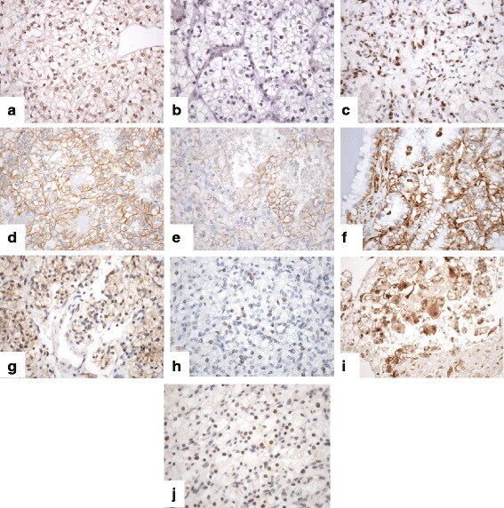
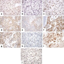
- Fig. 2 Immunohistochemical pattern in clear cell renal cell carcinomas displaying nuclear and/or cytoplasmic and/or membranous staining. a Snail, b Twist, c ZEB1, d beta-catenin, e E-cadherin, f Vimentin, g PTEN, h p110alpha, i p-Akt, j SETD2