Explore
 Validate
 Learn
Learn Western blot
 ELISA
ELISAAntibody data
- Antibody Data
- Antigen structure
- References [0]
- Comments [0]
- Validations
- Western blot [1]
- Immunohistochemistry [1]
Submit
Validation data
Reference
Comment
Report error
- Product number
- AM08433PU-N - Provider product page

- Provider
- Acris Antibodies GmbH
- Proper citation
- Acris Antibodies GmbH Cat#AM08433PU-N, RRID:AB_2035099
- Product name
- anti AKT1 / PKB pThr308
- Antibody type
- Monoclonal
- Antigen
- Synthetic peptide corresponding to residues surrounding T308 of human AKT1 protein
- Reactivity
- Human, Mouse
- Host
- Mouse
- Isotype
- IgG
- Antibody clone number
- 18F3.H11
- Vial size
- 0.1 mg
- Concentration
- 1,48 mg/ml (by UV absorbance at 280 nm)
No comments: Submit comment
Supportive validation
- Submitted by
- Acris Antibodies GmbH (provider)
- Main image
- Experimental details
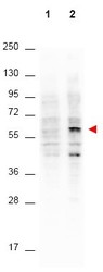
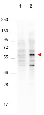
- Western blot using Anti-AKT pT308 monoclonal antibody shows detection of phosphorylated AKT (indicated by arrowhead at ~56 kDa) on PDGF stimulated NIH/3T3 cell lysates (lane 2). No reactivity is seen for non-phosphorylated AKT in untreated cells (lane 1). Each lane contained approximately 15 µg of lysate. All samples were loaded on to a 4-20% gradient gel for separation. After electrophoresis, the gel was blocked with 3% BSA in TBS for 30 min at RT. The membrane was probed with the primary antibody at a 1:4,000 dilution in TBS with 3% BSA, for 3 h at 4° C. For detection peroxidase conjugated Gt-a-Mouse IgG (Fc) was used at a 1:40,000 dilution for 1 h at 4° C. Data was captured using a Versadoc 4000 MP (Bio-Rad).
Supportive validation
- Submitted by
- Acris Antibodies GmbH (provider)
- Main image
- Experimental details
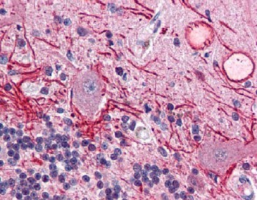
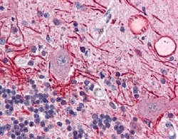
- Immunohistochemistry using Anti-AKT pT308 monoclonal antibody shows detection of phosphorylated AKT pT308 in human brain cerebellum tissue (40X). The antibody was used a dilution to 20 µg/mL. The image shows strong staining of Purkinje neurons and cell processes in the cerebellum. Staining was both cytosolic as well as occasionally nuclear, and the antibody showed minimal background staining. Tissue was formalin fixed and paraffin embedded. No pre-treatment of sample was required. The image shows the localization of antibody as the precipitated red signal, with a hematoxylin purple nuclear counterstain.