Explore
 Validate
 Learn
Learn Western blot
Antibody data
- Antibody Data
- Antigen structure
- References [3]
- Comments [0]
- Validations
- Western blot [1]
- Immunohistochemistry [1]
Submit
Validation data
Reference
Comment
Report error
- Product number
- MAB1919 - Provider product page

- Provider
- Abnova Corporation
- Proper citation
- Abnova Corporation Cat#MAB1919, RRID:AB_1678449
- Product name
- AKT1 (phospho S473) monoclonal antibody, clone 17B6.B11.A12.C3.G7.G12
- Antibody type
- Monoclonal
- Description
- Mouse monoclonal antibody raised against synthetic phosphopeptide of AKT1.
- Isotype
- IgG
- Antibody clone number
- 17B6.B11.A12.C3.G7.G12
- Storage
- Store at 4°C. For long term storage store at -20°C.Aliquot to avoid repeated freezing and thawing.
Submitted references Discovery of PDK1, one of the missing links in insulin signal transduction. Colworth Medal Lecture.
PKB/Akt: a key mediator of cell proliferation, survival and insulin responses?
Molecular cloning and identification of a serine/threonine protein kinase of the second-messenger subfamily.
Alessi DR
Biochemical Society transactions 2001 May;29(Pt 2):1-14
Biochemical Society transactions 2001 May;29(Pt 2):1-14
PKB/Akt: a key mediator of cell proliferation, survival and insulin responses?
Lawlor MA, Alessi DR
Journal of cell science 2001 Aug;114(Pt 16):2903-10
Journal of cell science 2001 Aug;114(Pt 16):2903-10
Molecular cloning and identification of a serine/threonine protein kinase of the second-messenger subfamily.
Jones PF, Jakubowicz T, Pitossi FJ, Maurer F, Hemmings BA
Proceedings of the National Academy of Sciences of the United States of America 1991 May 15;88(10):4171-5
Proceedings of the National Academy of Sciences of the United States of America 1991 May 15;88(10):4171-5
No comments: Submit comment
Supportive validation
- Submitted by
- Abnova Corporation (provider)
- Main image
- Experimental details
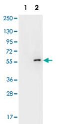
- Western blot using AKT1 (phospho S473) monoclonal antibody, clone 17B6.B11.A12.C3.G7.G12 (Cat # MAB1919) shows detection of phospho-AKT1 (indicated by arrowhead at ~56 KDa) on PDGF stimulated NIH/3T3 cell lysates (Lane 2).No reactivity is seen for non-phosphorylated AKT1 in untreated cells (Lane 1).Each lane contained approximately 10 ug of lysate.All samples were loaded on to a 4-20% gradient gel for separation.After electrophoresis, the gel was blocked with 5% BLOTTO in TBS for 90 min at RT.The membrane was probed with the primary antibody at a 1:10,000 dilution in TBS with 0.05% Tween-20 with 1% BSA, for 1 h at 4°C. For detection HRP conjugated Gt-a-Mouse IgG was used at a 1:20,000 dilution for 1 h at 4°C with FemtoMax™ enhanced chemiluminescent reagent.Images were captured using 2X2 binning for 10-20 sec using a BioSpectrum Imaging System (UVP Ltd.).
Supportive validation
- Submitted by
- Abnova Corporation (provider)
- Main image
- Experimental details
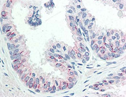
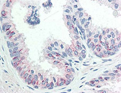
- Immunohistochemistry of formalin fixed and paraffin embedded using AKT1 (phospho S473) monoclonal antibody, clone 17B6.B11.A12.C3.G7.G12 (Cat # MAB1919) shows detection of Phospho-AKT1 S473 in human prostate tissue.The antibody was used at 20 ug/mL .The staining is much stronger than the weak basal level of phosphorylation in normal prostate tissue.Signal was developed using Dako's Techmate streptavidin-biotin reagents.Personal communication, Glenna Burmer, Lifespan Biosciences, Seattle, WA.
- Validation comment
- Immunohistochemistry (Formalin/PFA-fixed paraffin-embedded sections)