Explore
 Validate
 Learn
Learn Flow cytometry
Antibody data
- Antibody Data
- Antigen structure
- References [3]
- Comments [0]
- Validations
- Flow cytometry [1]
Submit
Validation data
Reference
Comment
Report error
- Product number
- 11-9715-41 - Provider product page

- Provider
- Invitrogen Antibodies
- Product name
- Anti-Phospho-AKT1 (Ser473) Monoclonal Antibody (SDRNR), FITC, eBioscience™
- Antibody type
- Monoclonal
- Antigen
- Other
- Description
- Description: This SDRNR monoclonal antibody recognizes human and mouse AKT (also known as Protein Kinase B (PKB)) when phosphorylated on S473. AKT is a serine/threonine protein kinase that plays a key role in multiple cellular processes including metabolism, proliferation, apoptosis/survival, and migration. There are three homologous isoforms of AKT: AKT1, AKT2, and AKT3. AKT is activated by binding of its pleckstrin homology (PH) domain to membrane phospholipids and by phosphorylation. Phosphorylation of AKT at T308 by PDK1 and at S473 is required for full activation of this kinase. AKT promotes cell survival by inhibiting apoptosis via phosphorylation and inactivation of several targets including Bad, Foxo1, c-Raf, and caspase-9. Deregulation of AKT has been implicated as a major contributing factor in many types of cancer. AKT is negatively regulated by the phosphatase PTEN as well as by the chemical inhibitor LY294002. Specificity of this SDRNR clone was determined by ELISA, flow cytometry, and western blotting. Applications Reported: This SDRNR antibody has been reported for use in intracellular staining followed by flow cytometric analysis. Applications Tested: This SDRNR antibody has been pre-titrated and tested by intracellular staining followed by flow cytometric analysis of normal human peripheral blood cells. This can be used at 5 µL (0.06 µg) per test. A test is defined as the amount (µg) of antibody that will stain a cell sample in a final volume of 100 µL. Cell number should be determined empirically but can range from 10^5 to 10^8 cells/test. Staining Protocol: All protocols work well for this monoclonal antibody. Use of Protocol A: Two-step protocol: intracellular (cytoplasmic) proteins allows for the greatest flexibility for detection of surface and intracellular (cytoplasmic) proteins. Use of Protocol B: One-step protocol: intracellular (nuclear) proteins is recommended for staining of transcription factors in conjunction with surface and phosphorylated intracellular (cytoplasmic) proteins. Protocol C: Two-step protocol: Fixation/Methanol allows for the greatest discrimination of phospho-specific signaling between unstimulated and stimulated samples, but with limitations on the ability to stain specific surface proteins (refer to "Clone Performance Following Fixation/Permeabilization" located in the Best Protocols Section under the Resources tab online). All Protocols can be found in the Flow Cytometry Protocols: "Staining Intracellular Antigens for Flow Cytometry Protocol" located in the Best Protocols Section under the Resources tab online. Excitation: 488 nm; Emission: 520 nm; Laser: Blue Laser. Filtration: 0.2 µm post-manufacturing filtered.
- Reactivity
- Human, Mouse
- Host
- Mouse
- Conjugate
- Green dye
- Isotype
- IgG
- Antibody clone number
- SDRNR
- Vial size
- 25 Tests
- Concentration
- 5 µL/Test
- Storage
- 4° C, store in dark, DO NOT FREEZE!
Submitted references Crosstalks between mTORC1 and mTORC2 variagate cytokine signaling to control NK maturation and effector function.
Store-Operated Ca2+ Entry Controls Clonal Expansion of T Cells through Metabolic Reprogramming.
Regulatory T Cell Migration Is Dependent on Glucokinase-Mediated Glycolysis.
Wang F, Meng M, Mo B, Yang Y, Ji Y, Huang P, Lai W, Pan X, You T, Luo H, Guan X, Deng Y, Yuan S, Chu J, Namaka M, Hughes T, Ye L, Yu J, Li X, Deng Y
Nature communications 2018 Nov 19;9(1):4874
Nature communications 2018 Nov 19;9(1):4874
Store-Operated Ca2+ Entry Controls Clonal Expansion of T Cells through Metabolic Reprogramming.
Vaeth M, Maus M, Klein-Hessling S, Freinkman E, Yang J, Eckstein M, Cameron S, Turvey SE, Serfling E, Berberich-Siebelt F, Possemato R, Feske S
Immunity 2017 Oct 17;47(4):664-679.e6
Immunity 2017 Oct 17;47(4):664-679.e6
Regulatory T Cell Migration Is Dependent on Glucokinase-Mediated Glycolysis.
Kishore M, Cheung KCP, Fu H, Bonacina F, Wang G, Coe D, Ward EJ, Colamatteo A, Jangani M, Baragetti A, Matarese G, Smith DM, Haas R, Mauro C, Wraith DC, Okkenhaug K, Catapano AL, De Rosa V, Norata GD, Marelli-Berg FM
Immunity 2017 Nov 21;47(5):875-889.e10
Immunity 2017 Nov 21;47(5):875-889.e10
No comments: Submit comment
Supportive validation
- Submitted by
- Invitrogen Antibodies (provider)
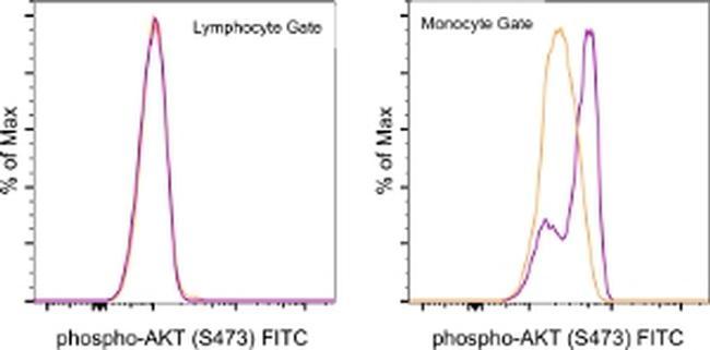
- Main image
- Experimental details
- Normal human peripheral blood cells were unstimulated (orange histogram) or stimulated with Lipopolysaccharide (LPS) Solution (500X) (Product # 00-4976-03) (purple histogram), then intracellularly stained with Anti-Human/Mouse phospho-AKT (S473) FITC using the Intracellular Fixation and Permeabilization Buffer Set (Product # 88-8824-00) and protocol. Cell in the lymphocyte (left) and monocyte (right) gates were used for analysis.
- Conjugate
- Green dye