Explore
 Validate
 Learn
Learn Flow cytometry
Antibody data
- Antibody Data
- Antigen structure
- References [10]
- Comments [0]
- Validations
- Flow cytometry [1]
- Other assay [9]
Submit
Validation data
Reference
Comment
Report error
- Product number
- 11-9715-42 - Provider product page

- Provider
- Invitrogen Antibodies
- Product name
- Phospho-AKT1 (Ser473) Monoclonal Antibody (SDRNR), FITC, eBioscience™
- Antibody type
- Monoclonal
- Antigen
- Other
- Description
- Description: This SDRNR monoclonal antibody recognizes human and mouse AKT (also known as Protein Kinase B (PKB)) when phosphorylated on S473. AKT is a serine/threonine protein kinase that plays a key role in multiple cellular processes including metabolism, proliferation, apoptosis/survival, and migration. There are three homologous isoforms of AKT: AKT1, AKT2, and AKT3. AKT is activated by binding of its pleckstrin homology (PH) domain to membrane phospholipids and by phosphorylation. Phosphorylation of AKT at T308 by PDK1 and at S473 is required for full activation of this kinase. AKT promotes cell survival by inhibiting apoptosis via phosphorylation and inactivation of several targets including Bad, Foxo1, c-Raf, and caspase-9. Deregulation of AKT has been implicated as a major contributing factor in many types of cancer. AKT is negatively regulated by the phosphatase PTEN as well as by the chemical inhibitor LY294002. Specificity of this SDRNR clone was determined by ELISA, flow cytometry, and western blotting. Applications Reported: This SDRNR antibody has been reported for use in intracellular staining followed by flow cytometric analysis. Applications Tested: This SDRNR antibody has been pre-titrated and tested by intracellular staining followed by flow cytometric analysis of normal human peripheral blood cells. This can be used at 5 µL (0.06 µg) per test. A test is defined as the amount (µg) of antibody that will stain a cell sample in a final volume of 100 µL. Cell number should be determined empirically but can range from 10^5 to 10^8 cells/test. Staining Protocol: All protocols work well for this monoclonal antibody. Use of Protocol A: Two-step protocol: intracellular (cytoplasmic) proteins allows for the greatest flexibility for detection of surface and intracellular (cytoplasmic) proteins. Use of Protocol B: One-step protocol: intracellular (nuclear) proteins is recommended for staining of transcription factors in conjunction with surface and phosphorylated intracellular (cytoplasmic) proteins. Protocol C: Two-step protocol: Fixation/Methanol allows for the greatest discrimination of phospho-specific signaling between unstimulated and stimulated samples, but with limitations on the ability to stain specific surface proteins (refer to "Clone Performance Following Fixation/Permeabilization" located in the Best Protocols Section under the Resources tab online). All Protocols can be found in the Flow Cytometry Protocols: "Staining Intracellular Antigens for Flow Cytometry Protocol" located in the Best Protocols Section under the Resources tab online. Excitation: 488 nm; Emission: 520 nm; Laser: Blue Laser. Filtration: 0.2 µm post-manufacturing filtered.
- Reactivity
- Human, Mouse
- Host
- Mouse
- Conjugate
- Green dye
- Isotype
- IgG
- Antibody clone number
- SDRNR
- Vial size
- 100 Tests
- Concentration
- 5 µL/Test
- Storage
- 4° C, store in dark, DO NOT FREEZE!
Submitted references Multicellular immune dynamics implicate PIM1 as a potential therapeutic target for uveitis.
Combinatorial immunotherapy of N-803 (IL-15 superagonist) and dinutuximab with ex vivo expanded natural killer cells significantly enhances in vitro cytotoxicity against GD2(+) pediatric solid tumors and in vivo survival of xenografted immunodeficient NSG mice.
Novel cytokine-antibody fusion protein, N-820, to enhance the functions of ex vivo expanded natural killer cells against Burkitt lymphoma.
Long-Term Programming of CD8 T Cell Immunity by Perinatal Exposure to Glucocorticoids.
IL-15 negatively regulates curdlan-induced IL-23 production by human monocyte-derived dendritic cells and subsequent Th17 response.
Fc Receptor-Like 1 as a Promising Target for Immunotherapeutic Interventions of B-Cell-Related Disorders.
Crosstalks between mTORC1 and mTORC2 variagate cytokine signaling to control NK maturation and effector function.
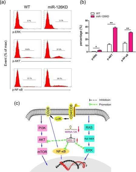
MicroRNA-126 deficiency enhanced the activation and function of CD4(+) T cells by elevating IRS-1 pathway.
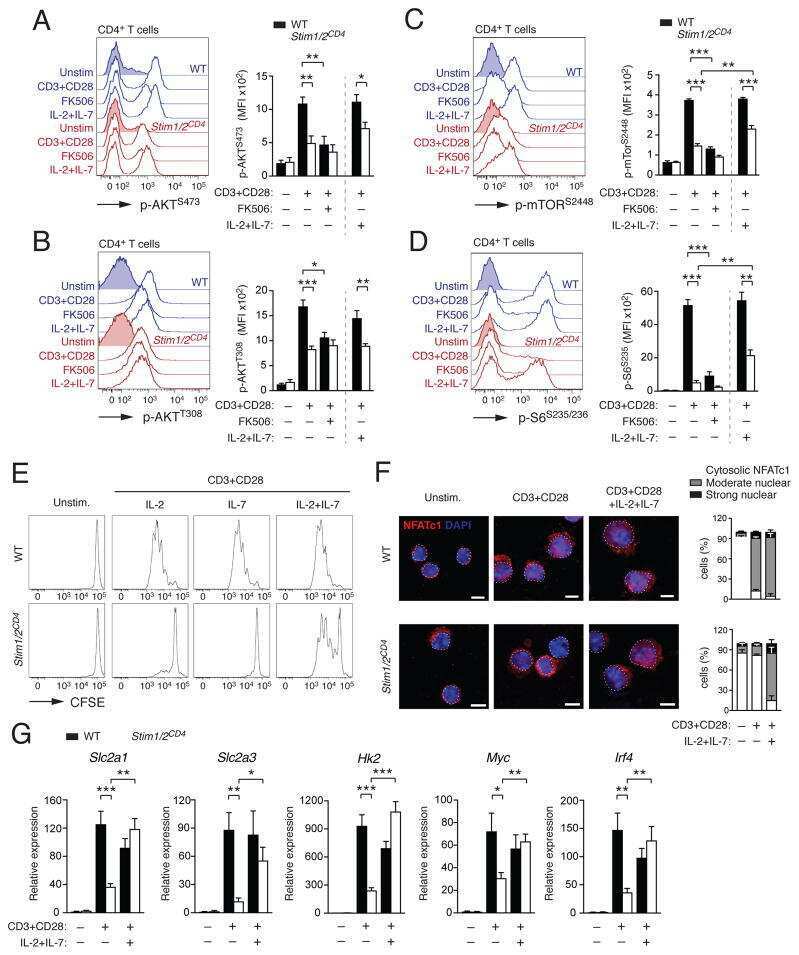
Store-Operated Ca(2+) Entry Controls Clonal Expansion of T Cells through Metabolic Reprogramming.
Regulatory T Cell Migration Is Dependent on Glucokinase-Mediated Glycolysis.
Li H, Xie L, Zhu L, Li Z, Wang R, Liu X, Huang Z, Chen B, Gao Y, Wei L, He C, Ju R, Liu Y, Liu X, Zheng Y, Su W
Nature communications 2022 Oct 4;13(1):5866
Nature communications 2022 Oct 4;13(1):5866
Combinatorial immunotherapy of N-803 (IL-15 superagonist) and dinutuximab with ex vivo expanded natural killer cells significantly enhances in vitro cytotoxicity against GD2(+) pediatric solid tumors and in vivo survival of xenografted immunodeficient NSG mice.
Chu Y, Nayyar G, Jiang S, Rosenblum JM, Soon-Shiong P, Safrit JT, Lee DA, Cairo MS
Journal for immunotherapy of cancer 2021 Jul;9(7)
Journal for immunotherapy of cancer 2021 Jul;9(7)
Novel cytokine-antibody fusion protein, N-820, to enhance the functions of ex vivo expanded natural killer cells against Burkitt lymphoma.
Chu Y, Nayyar G, Kham Su N, Rosenblum JM, Soon-Shiong P, Lee J, Safrit JT, Barth M, Lee D, Cairo MS
Journal for immunotherapy of cancer 2020 Oct;8(2)
Journal for immunotherapy of cancer 2020 Oct;8(2)
Long-Term Programming of CD8 T Cell Immunity by Perinatal Exposure to Glucocorticoids.
Hong JY, Lim J, Carvalho F, Cho JY, Vaidyanathan B, Yu S, Annicelli C, Ip WKE, Medzhitov R
Cell 2020 Mar 5;180(5):847-861.e15
Cell 2020 Mar 5;180(5):847-861.e15
IL-15 negatively regulates curdlan-induced IL-23 production by human monocyte-derived dendritic cells and subsequent Th17 response.
Eken A, Okus Z, Erdem S, Azizoglu ZB, Haliloglu Y, Bicer A, Gur TN, Yilmaz E, Karakukcu M, Altuntas HD, Canatan H
Northern clinics of Istanbul 2019;6(4):379-387
Northern clinics of Istanbul 2019;6(4):379-387
Fc Receptor-Like 1 as a Promising Target for Immunotherapeutic Interventions of B-Cell-Related Disorders.
Yousefi Z, Sharifzadeh S, Yar-Ahmadi V, Andalib A, Eskandari N
Biomarker insights 2019;14:1177271919882351
Biomarker insights 2019;14:1177271919882351
Crosstalks between mTORC1 and mTORC2 variagate cytokine signaling to control NK maturation and effector function.
Wang F, Meng M, Mo B, Yang Y, Ji Y, Huang P, Lai W, Pan X, You T, Luo H, Guan X, Deng Y, Yuan S, Chu J, Namaka M, Hughes T, Ye L, Yu J, Li X, Deng Y
Nature communications 2018 Nov 19;9(1):4874
Nature communications 2018 Nov 19;9(1):4874
MicroRNA-126 deficiency enhanced the activation and function of CD4(+) T cells by elevating IRS-1 pathway.
Chu F, Hu Y, Zhou Y, Guo M, Lu J, Zheng W, Xu H, Zhao J, Xu L
Clinical and experimental immunology 2018 Feb;191(2):166-179
Clinical and experimental immunology 2018 Feb;191(2):166-179
Store-Operated Ca(2+) Entry Controls Clonal Expansion of T Cells through Metabolic Reprogramming.
Vaeth M, Maus M, Klein-Hessling S, Freinkman E, Yang J, Eckstein M, Cameron S, Turvey SE, Serfling E, Berberich-Siebelt F, Possemato R, Feske S
Immunity 2017 Oct 17;47(4):664-679.e6
Immunity 2017 Oct 17;47(4):664-679.e6
Regulatory T Cell Migration Is Dependent on Glucokinase-Mediated Glycolysis.
Kishore M, Cheung KCP, Fu H, Bonacina F, Wang G, Coe D, Ward EJ, Colamatteo A, Jangani M, Baragetti A, Matarese G, Smith DM, Haas R, Mauro C, Wraith DC, Okkenhaug K, Catapano AL, De Rosa V, Norata GD, Marelli-Berg FM
Immunity 2017 Nov 21;47(5):875-889.e10
Immunity 2017 Nov 21;47(5):875-889.e10
No comments: Submit comment
Supportive validation
- Submitted by
- Invitrogen Antibodies (provider)
- Main image
- Experimental details
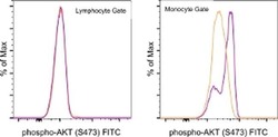
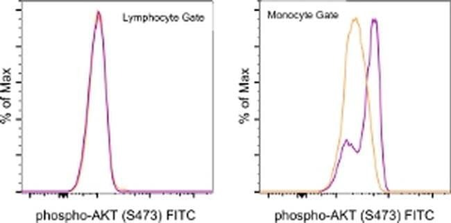
- Normal human peripheral blood cells were unstimulated (orange histogram) or stimulated with Lipopolysaccharide (LPS) Solution (500X) (Product # 00-4976-03) (purple histogram), then intracellularly stained with Anti-Human/Mouse phospho-AKT (S473) FITC using the Intracellular Fixation and Permeabilization Buffer Set (Product # 88-8824-00) and protocol. Cell in the lymphocyte (left) and monocyte (right) gates were used for analysis.
- Conjugate
- Green dye
Supportive validation
- Submitted by
- Invitrogen Antibodies (provider)
- Main image
- Experimental details
- NULL
- Conjugate
- Green dye
- Submitted by
- Invitrogen Antibodies (provider)
- Main image

- Experimental details
- NULL
- Conjugate
- Green dye
- Submitted by
- Invitrogen Antibodies (provider)
- Main image
- Experimental details
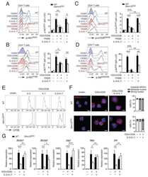
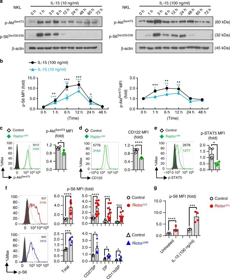
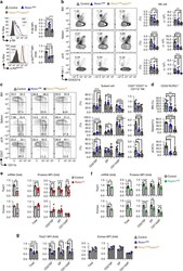
- Fig. 4 Crosstalk between mTORC1 and mTORC2 during NK cell activation. a Immunoblotting to detect the phosphorylation (p-) of S6 at ser235/236 (p-S6 ser235/236 ) and Akt at Ser473 (p-Akt ser473 ) in NKL cells after stimulation with IL-15 (10 ng/ml) (left) or IL-15 (100 ng/ml) (right) for the indicated time periods. beta-Actin was used as an internal control. The data shown are representative of three independent experiments. Uncropped gels are shown in Supplementary Fig. 8 . b Intracellular flow cytometric analysis of p-S6 ser235/236 and p-Akt ser473 in murine splenic NK cells (CD3 - CD19 - NK1.1 + ) after stimulation with IL-15 (100 or 10 ng/ml) for the indicated time periods. The MFI of p-S6 ser235/236 or p-Akt ser473 at each time point was normalized to the baseline reading at the 0-h time point for each individual mouse. Four independent individual mice were used. c - e Flow cytometric analysis and cumulative results for p-Akt Ser473 ( c ), CD122 ( d ), and p-STAT5 Y694 ( e ) in splenic NK cells (CD3 - CD19 - NK1.1 + ) from control versus Raptor NK mice. f Intracellular flow cytometric analysis and cumulative results for p-S6 ser235/236 in total splenic NK cells (CD3 - CD19 - NK1.1 + ) and the indicated subpopulations thereof from control versus Rictor / (top) or control versus Rictor NK (bottom) mice. g Statistical quantification of p-S6 ser235/236 (MFI relative to the untreated control littermates) in splenic NK cells (CD3 - CD19 - NK1.1 + ) wit
- Conjugate
- Green dye
- Submitted by
- Invitrogen Antibodies (provider)
- Main image
- Experimental details
- Fig. 5 mTORC2 and mTORC1 promote NK cell maturation by controlling the expression of Tbx21 and Eomes in a cooperative and nonredundant manner. a Intracellular flow cytometric analysis and cumulative results for the phosphorylation (p-) of S6 at ser235/236 (p-S6 ser235/236 ) (top) and Akt at Ser473 (p-Akt Ser473 ) (bottom) in splenic NK cells (CD3 - CD19 - NK1.1 + ) from mice of the indicated genotype. b Cumulative ratio and enumeration of NK cells (CD3 - CD19 - NK1.1 + ) in the BM, spleen, and peripheral lymph nodes (pLNs) from mice of the indicated genotype. c Flow cytometric analysis and cumulative frequencies of subpopulations of NK cells (CD3 - CD19 - NK1.1 + NKp46 + ) in the BM, spleen, and pLNs (left) and the calculated ratio of CD27 - versus CD27 + cells among CD11b + NK cells (right). d The cumulative frequencies depicting the CD43 + KLRG1 + subset of NK cells (CD3 - CD19 - NK1.1 + NKp46 + ) in the BM, spleen, and pLNs from mice of the indicated genotype. e , f Tbx21 and Eomes mRNA and protein expression in control versus Rictor / mice ( e ) and control versus Raptor NK mice ( f ), as assessed by quantitative RT-PCR and flow cytometry, respectively. Purified splenic NK cells were used for quantitative RT-PCR (left). Cumulative data for Tbx21 and Eomes protein expression in total splenic NK cells (CD3 - CD19 - NK1.1 + NKp46 + ) and the indicated subpopulations thereof were analyzed by flow cytometry (right). g Cumulative data for Tbx21 and Eomes pr
- Conjugate
- Green dye
- Submitted by
- Invitrogen Antibodies (provider)
- Main image

- Experimental details
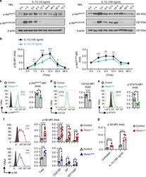
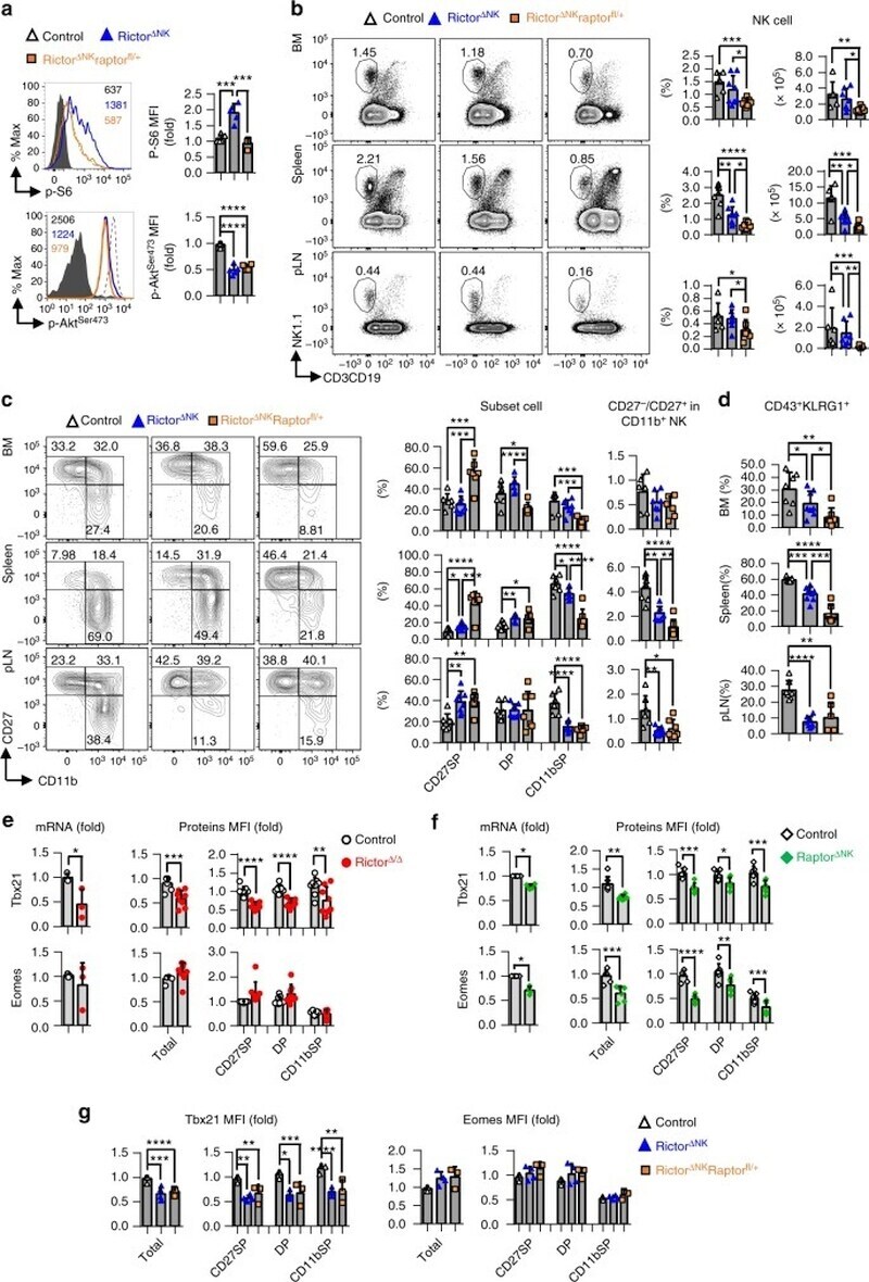
- FIGURE 3 IL-15 exposure downregulates surface Dectin-1 receptor expression and impacts signalling in DCs (A) After five days of culture in with IL-4/GM-CSF or IL-4/GM-CSF/IL-15 DCs were surface stained for Clec7 (Dectin-1) and a representative flow graph and (B) quantification of mean fluorescent intensity (MFI) as bar graph are presented. The experiment was run in triplicate wells and a representative result from a single donor was shown, the experiment was repeated with four different donors. (C) DCs from ""A"" were stimulated with curdlan (50 ug/ml) and phosphorylation of p38, AKT, SRC, NFKB p65, IkBa, ERK1/2 and IRAK4 was measured by phospho-flow assay. A representative result from PBMCs of a single donor was charted. *p
- Conjugate
- Green dye
- Submitted by
- Invitrogen Antibodies (provider)
- Main image
- Experimental details
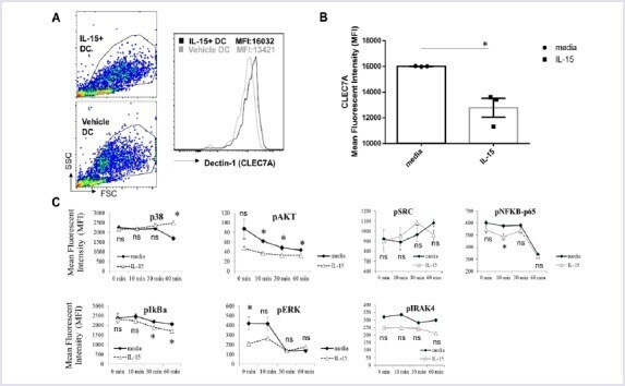
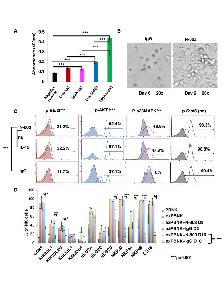
- Figure 1 N-803 increased the viability and proliferation of exPBNK with enhanced p-Stat3, p-Stat5, pAKT, p-p38MAPK and NK activating receptors. PBMNCs were stimulated with irradiated genetically modified K562-mbIL21-41BBL cells for 2-3 weeks. (A) Purified exPBNK cells were cultured in complete medium with 0.35 ng/mL (low) or 3.5 ng/mL (high) N-803 or molar equivalent dose of IgG for 3 days. NK viability and proliferation were monitored by MTS assays. The amount of 490 nm absorbance is directly proportional to the number of living exPBNK cells in the culture. The exPBNK cells with N-803 at 0.35 ng/mL or 3.5 ng/mL have significantly higher viability as compared with IgG or medium controls (p
- Conjugate
- Green dye
- Submitted by
- Invitrogen Antibodies (provider)
- Main image
- Experimental details
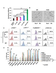
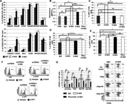
- Figure 1 N-820 enhanced the expression of NK activating receptors and proliferation of exPBNK with enhanced Ki67, p-Stat5, and CD25 levels. The percentages of NK activating and inhibitory receptors on exPBNK surface were monitored by flow cytometry analysis at day 3 (A). The NK proliferation was monitored by CellTiter 96 AQueous One solution cell proliferation assay (Promega) according to the manufacturer's instruction (B). Intracellular Ki67 (C), Phosphorylated STAT5 (p-STAT5) (D) and phosphorylated Akt (p-AKT) (E) were monitored by flow cytometry analysis at day 3. Ki67 (p
- Conjugate
- Green dye
- Submitted by
- Invitrogen Antibodies (provider)
- Main image
- Experimental details
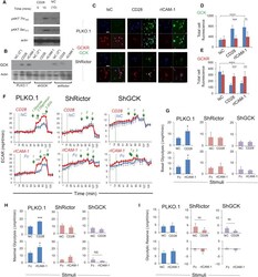
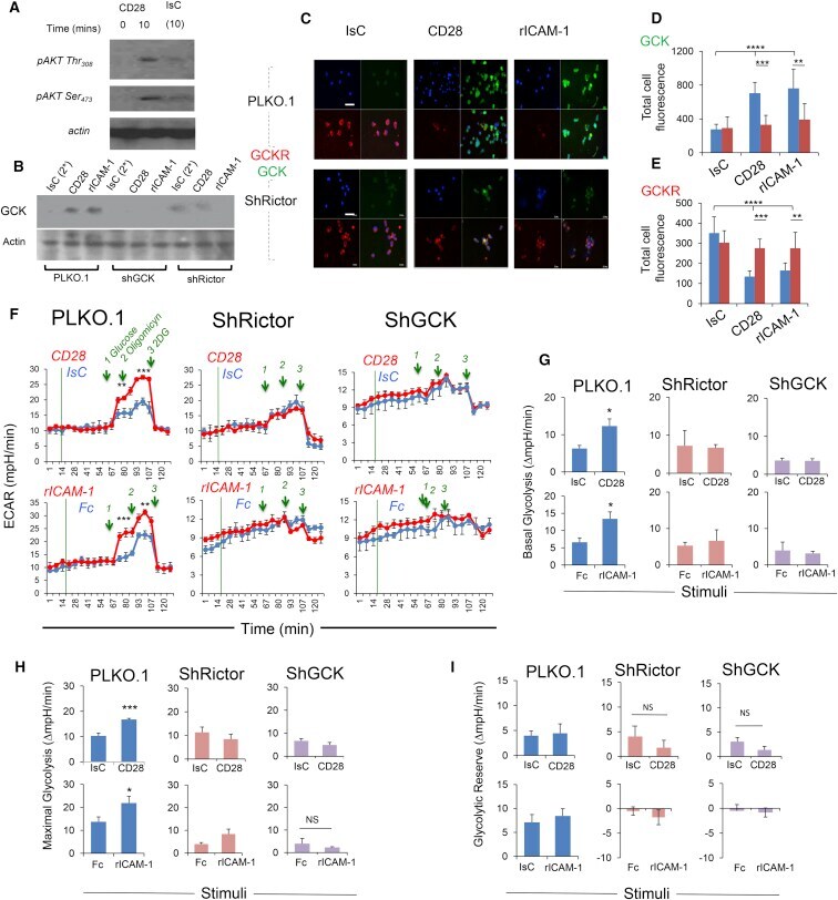
- Figure 5 mTORC2 Controls Metabolic Reprogramming Induced by Pro-migratory Stimuli (A) Phosphorylation of AKT at Thr 308 and Ser 473 in Treg cells activated with CD28- or IsC-antibody ligation was measured by immunoblotting. (B) Treg cells were virally transfected with Rictor-specific or GCK-specific or non-sense (PLKO.1) sh-RNAs, as described in STAR Methods . Expression of GCK was measured by immunoblotting 24 hr later. (C-E) Expression of GCK and GCKR by control or Rictor-deficient Treg cells following CD28 or LFA-1 activation for 45 min. Bar graphs (D and E) show the mean protein expression (Total cell fluorescence) measured in 3 independent experiments by ImageJ software +- SD. Scale bar, 40 mum. (F-I) ECAR of CD28- or LFA-1-stimulated Rictor- and GCK-deficient and control T cells was measured with an extracellular flux analyzer. A glycolysis stress assay was performed by adding the indicated compounds at the time points indicated by the green lines. The basal and maximal glycolysis and the glycolytic reserve are shown in (G), (H), and (I), respectively (+-SEM). N = 2. * p < 0.05 *** p < 0.005; **** p < 0.001. Please see also Figure S5 .
- Conjugate
- Green dye
- Submitted by
- Invitrogen Antibodies (provider)
- Main image
- Experimental details
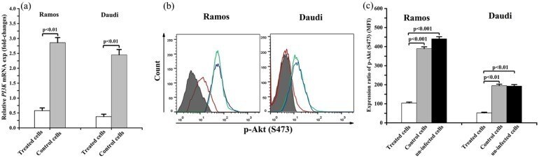
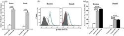
- Figure 4. The effect of FCRL1 knockdown the extent of the expression of PI3K /p-AKT pathway in the Ramos and Daudi cells.(a) The real-time PCR approach revealed a significant decrease in theextent of the PI3K gene expression in the BL cellsafter 3 days of the infection procedure. The extent of the expression ofthe p-AKT protein was measured by (b) cytometry and (c) using the FlowJosoftware on day 4 of infection procedure. Shaded-matched representsmatched isotype control antibody, blue represents uninfected cells,green represents control cells, and red represents treated cells. Dataare shown as mean +- standard deviation. BL indicates Burkitt lymphoma; FCRL, Fc receptor-like; PCR, polymerasechain reaction.
- Conjugate
- Green dye