 Explore
 Validate
 Learn
Learn Western blot
Antibody data
- Antibody Data
- Antigen structure
- References [1]
- Comments [0]
- Validations
- Western blot [2]
- Immunocytochemistry [3]
- Immunohistochemistry [2]
Submit
Validation data
Reference
Comment
Report error
- Product number
- 710005 - Provider product page

- Provider
- Invitrogen Antibodies
- Product name
- AKT1 Recombinant Polyclonal Antibody (43HCLC)
- Antibody type
- Polyclonal
- Antigen
- Other
- Description
- This antibody is predicted to react with mouse based on sequence homology.
- Antibody clone number
- 43HCLC
- Concentration
- 0.5 mg/mL
Submitted references Crocin Inhibits Oxidative Stress and Pro-inflammatory Response of Microglial Cells Associated with Diabetic Retinopathy Through the Activation of PI3K/Akt Signaling Pathway.
Yang X, Huo F, Liu B, Liu J, Chen T, Li J, Zhu Z, Lv B
Journal of molecular neuroscience : MN 2017 Apr;61(4):581-589
Journal of molecular neuroscience : MN 2017 Apr;61(4):581-589
No comments: Submit comment
Supportive validation
- Submitted by
- Invitrogen Antibodies (provider)
- Main image

- Experimental details
- Western blot analysis of AKT in MCF-7 cell lysate (30 µg/lane) using an AKT Recombinant Rabbit Polyclonal Antibody (Product # 710005) at a dilution of 2.5 µg/mL. NBT/BCIP was used as the substrate (Product # WB7105).

- Submitted by
- Invitrogen Antibodies (provider)
- Main image

- Experimental details
- Western blot analysis of AKT was performed by loading 20 µg of K562, HEK-293, U-2 OS, A549, MCF-7, A431 and PC3 cell lysates using Novex®NuPAGE® 4-12% Bis-Tris gel (Product # NP0321BOX), XCell SureLock Electrophoresis System (Product # EI0002), Novex® Sharp Pre-Stained Protein Standard (Product # LC5800), and iBlot® Dry Blotting System (Product # IB21001). Proteins were transferred to a nitrocellulose membrane and blocked with 5% skim milk for 1 hour at room temperature. AKT was detected at ~56 kDa using AKT Recombinant Rabbit Polyclonal Antibody (Product # 710005) at a 1:1000 dilution in 2.5% skim milk at 4°C overnight on a rocking platform. Detection was performed using an HRP-conjugated Goat anti-Rabbit secondary antibody (Product # G-21234) at a 1:5000 dilution and chemiluminescent detection was performed using Pierce™ ECL Western blotting Substrate (Product # 32106).

Supportive validation
- Submitted by
- Invitrogen Antibodies (provider)
- Main image

- Experimental details
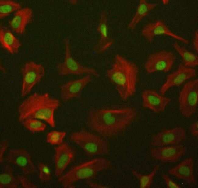
- Immunofluorescent analysis of AKT in HeLa cells using an AKT Recombinant Rabbit Polyclonal Antibody (Product # 710005) at a dilution of 5 µg/mL followed by detection using an Alexa Fluor 488-conjugated Goat anti-Rabbit secondary antibody at a dilution of 1:1000 (green) and Alexa Fluor 594 Phalloidin (red).

- Submitted by
- Invitrogen Antibodies (provider)
- Main image

- Experimental details
- Immunofluorescent analysis of AKT in HeLa cells using an AKT Recombinant Rabbit Polyclonal Antibody (Product # 710005) at a dilution of 5 µg/mL followed by detection using an Alexa Fluor 488-conjugated Goat anti-Rabbit secondary antibody at a dilution of 1:1000 (green) and Alexa Fluor 594 Phalloidin (red).
- Submitted by
- Invitrogen Antibodies (provider)
- Main image
- Experimental details
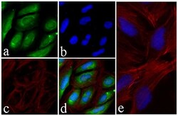
- Immunofluorescent analysis of AKT was performed on 70% confluent log phase U-2 OS cells. The cells were fixed with 4% paraformaldehyde for 15 minutes, permeabilized with 0. 25% Triton X-100 for 10 minutes, and blocked with 5% BSA for 1 hour at room temperature. The cells were labeled with AKT Recombinant Rabbit Polyclonal Antibody (Product # 710005) at a dilution of 1:500 in 1% BSA and incubated for 3 hours at room temperature and then labeled with Alexa Fluor® 488 Goat anti-Rabbit IgG secondary antibody (Product # A-11008) at a dilution of 1:400 for 30 minutes at room temperature (Panel a: green). Nuclei (Panel b: blue) were stained with SlowFade® Gold Antifade Mountant with DAPI (Product # S36938). F-actin (Panel c: red) was stained with Alexa Fluor® 594 phalloidin (Product # A12381). Panel d is a merged image showing cytoplasmic and nuclear localization. Panel e is a control without primary antibody. The images were captured using a Nikon microscope at 20X magnification.

Supportive validation
- Submitted by
- Invitrogen Antibodies (provider)
- Main image

- Experimental details
- Immunohistochemistry analysis of AKT showing staining in the cytoplasm of paraffin-embedded human prostate carcinoma (right) compared to a negative control without primary antibody (left). To expose target proteins, antigen retrieval was performed using 10 mM sodium citrate (pH 6.0), microwaved for 8-15 min. Following antigen retrieval, tissues were blocked in 3% H2O2-methanol for 15 min at room temperature, washed with ddH2O and PBS, and then probed with AKT Recombinant Rabbit Polyclonal Antibody (Product # 710005) diluted in 3% BSA-PBS at a dilution of 1:50 overnight at 4°C in a humidified chamber. Tissues were washed extensively in PBST and detection was performed using a HRP-conjugated secondary antibody followed by colorimetric detection using a DAB kit. Tissues were counterstained with hematoxylin and dehydrated with ethanol and xylene to prep for mounting.

- Submitted by
- Invitrogen Antibodies (provider)
- Main image

- Experimental details
- Immunohistochemistry analysis of AKT showing staining in the cytoplasm of paraffin-embedded mouse prostate tissue (right) compared to a negative control without primary antibody (left). To expose target proteins, antigen retrieval was performed using 10 mM sodium citrate (pH 6.0), microwaved for 8-15 min. Following antigen retrieval, tissues were blocked in 3% H2O2-methanol for 15 min at room temperature, washed with ddH2O and PBS, and then probed with AKT Recombinant Rabbit Polyclonal Antibody (Product # 710005) diluted in 3% BSA-PBS at a dilution of 1:50 overnight at 4°C in a humidified chamber. Tissues were washed extensively in PBST and detection was performed using a HRP-conjugated secondary antibody followed by colorimetric detection using a DAB kit. Tissues were counterstained with hematoxylin and dehydrated with ethanol and xylene to prep for mounting.
