 Explore
 Validate
 Learn
Learn Western blot
Antibody data
- Antibody Data
- Antigen structure
- References [2]
- Comments [0]
- Validations
- Western blot [3]
- Immunocytochemistry [2]
- Other assay [1]
Submit
Validation data
Reference
Comment
Report error
- Product number
- 710122 - Provider product page

- Provider
- Invitrogen Antibodies
- Product name
- Phospho-AKT1 (Thr308) Recombinant Polyclonal Antibody (B18HCLC)
- Antibody type
- Polyclonal
- Antigen
- Synthetic peptide
- Description
- Recombinant rabbit polyclonal antibodies are unique offerings from Thermo Fisher Scientific. They are comprised of a selection of multiple different recombinant monoclonal antibodies, providing the best of both worlds - the sensitivity of polyclonal antibodies with the specificity of monoclonal antibodies - all delivered with the consistency only found in a recombinant antibody. While functionally the same as a polyclonal antibody - recognizing multiple epitope sites on the target and producing higher detection sensitivity for low abundance targets - a recombinant rabbit polyclonal antibody has a known mixture of light and heavy chains. The exact population can be produced in every lot, circumventing the biological variability typically associated with polyclonal antibody production.
- Reactivity
- Human, Mouse
- Host
- Rabbit
- Isotype
- IgG
- Antibody clone number
- B18HCLC
- Vial size
- 100 µg
- Concentration
- 0.5 mg/mL
- Storage
- Store at 4°C short term. For long term storage, store at -20°C, avoiding freeze/thaw cycles.
Submitted references CDKL3 promotes osteosarcoma progression by activating Akt/PKB.
Neonatal maternal deprivation impairs localized de novo activity-induced protein translation at the synapse in the rat hippocampus.
He A, Ma L, Huang Y, Zhang H, Duan W, Li Z, Fei T, Yuan J, Wu H, Liu L, Bai Y, Dai W, Wang Y, Li H, Sun Y, Wang Y, Wang C, Yuan T, Yang Q, Tian S, Dong M, Sheng R, Xiang D
Life science alliance 2020 May;3(5)
Life science alliance 2020 May;3(5)
Neonatal maternal deprivation impairs localized de novo activity-induced protein translation at the synapse in the rat hippocampus.
Ahmad F, Salahuddin M, Alsamman K, Herzallah HK, Al-Otaibi ST
Bioscience reports 2018 Jun 29;38(3)
Bioscience reports 2018 Jun 29;38(3)
No comments: Submit comment
Supportive validation
- Submitted by
- Invitrogen Antibodies (provider)
- Main image

- Experimental details
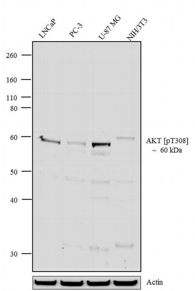
- Western blot analysis was performed on whole cell extracts (20 µg lysate) of LNCaP (Lane 1), PC-3 (Lane 2), U-87 MG (lane 3) and NIH/3T3 (lane 4). The blots were probed with Recombinant Rabbit Polyclonal Anti-AKT (pT308) Antibody (Product # 710122, 0.5-2 µg/mL) and detected by chemiluminescence using Goat anti-Rabbit IgG (H+L) Superclonal™ Secondary Antibody, HRP conjugate (Product # A27036, 0.4 µg/mL, 1:2500 dilution). A 60 kDa band corresponding to AKT (pT308) was observed across cell lines tested. Known quantity of protein samples were electrophoresed using Novex® NuPAGE® 4-12 % Bis-Tris gel (Product # NP0321BOX), XCell SureLock™ Electrophoresis System (Product # EI0002) and Novex® Sharp Pre-Stained Protein Standard (Product # LC5800). Resolved proteins were then transferred onto a nitrocellulose membrane with iBlot® 2 Dry Blotting System (Product # IB21001). The membrane was probed with the relevant primary and secondary Antibody following blocking with 5 % BSA. Chemiluminescent detection was performed using Pierce™ ECL Western blotting Substrate (Product # 32106).
- Submitted by
- Invitrogen Antibodies (provider)
- Main image

- Experimental details
- Western blot analysis was performed on whole cell extracts (20 µg lysate) of LNCaP (Lane 1), PC-3 (Lane 2), U-87 MG (lane 3) and NIH/3T3 (lane 4). The blots were probed with Recombinant Rabbit Polyclonal Anti-AKT (pT308) Antibody (Product # 710122, 0.5-2 µg/mL) and detected by chemiluminescence using Goat anti-Rabbit IgG (H+L) Superclonal™ Secondary Antibody, HRP conjugate (Product # A27036, 0.4 µg/mL, 1:2500 dilution). A 60 kDa band corresponding to AKT (pT308) was observed across cell lines tested. Known quantity of protein samples were electrophoresed using Novex® NuPAGE® 4-12 % Bis-Tris gel (Product # NP0321BOX), XCell SureLock™ Electrophoresis System (Product # EI0002) and Novex® Sharp Pre-Stained Protein Standard (Product # LC5800). Resolved proteins were then transferred onto a nitrocellulose membrane with iBlot® 2 Dry Blotting System (Product # IB21001). The membrane was probed with the relevant primary and secondary Antibody following blocking with 5 % BSA. Chemiluminescent detection was performed using Pierce™ ECL Western blotting Substrate (Product # 32106).

- Submitted by
- Invitrogen Antibodies (provider)
- Main image
- Experimental details
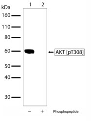
- Western blot analysis of Phospho-AKT pThr308 in whole cell extracts of U87-MG using a Phospho-AKT pThr308 Recombinant Rabbit Polyclonal Antibody (Product # 710122) at a dilution of 1 µg/mL. To confirm specificity, competition was performed by preincubation with the phosphopeptide to inhibit antibody binding (lane 2). Results show a band at ~60kDa.

Supportive validation
- Submitted by
- Invitrogen Antibodies (provider)
- Main image
- Experimental details
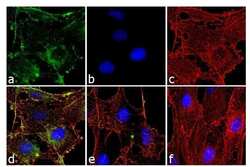
- Immunofluorescence analysis Phospho-AKT pThr308 was done on 70% confluent log phase NIH/3T3 cells treated with 50 ng of PDGF for 10 minutes. The cells were fixed with 4% paraformaldehyde for 10 minutes, permeabilized with 0.1% Triton™ X-100 for 10 minutes, and blocked with 1% BSA for 1 hour at room temperature. The cells were labeled with of Phospho-AKT pThr308 (B18HCLC), Recombinant Rabbit Polyclonal Antibody (Product # 710122) at 2 µg/mL in 0.1% BSA and incubated for 3 hours at room temperature and then labeled with Goat anti-Rabbit IgG (H+L) Superclonal™ Secondary Antibody, Alexa Fluor® 488 conjugate (Product # A27034) at a dilution of 1:2000 for 45 minutes at room temperature (Panel a: green). Nuclei (Panel b: blue) were stained with SlowFade® Gold Antifade Mountant with DAPI (Product # S36938). F-actin (Panel c: red) was stained with Alexa Fluor® 555 Rhodamine Phalloidin (Product # R415, 1:300). Panel d is a merged image showing cytoplasmic and membranous localization. Panel e is untreated cells with no signal. Panel f is a no primary antibody control. The images were captured at 60X magnification.

- Submitted by
- Invitrogen Antibodies (provider)
- Main image

- Experimental details
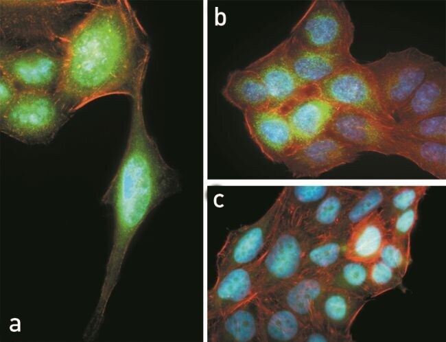
- Immunofluorescent analysis of Phospho-AKT pThr308 in HeLa cells using a Phospho-AKT pThr308 Recombinant Rabbit Polyclonal Antibody (Product # 710122) followed by detection using an Alexa Fluor 488-conjugated Goat anti-Rabbit secondary antibody (green), nuclei staining using DAPI (blue) and actin staining using Alexa Fluor 594 phalloidin (red). Images an And B are a composite image showing showing nuclear localization of phosphorylated AKT and Image C shows the results of a competition assay using phospho AKT (pT308) peptide.
Supportive validation
- Submitted by
- Invitrogen Antibodies (provider)
- Main image

- Experimental details
- Figure 6. Cyclin-dependent kinase-like 3 (CDKL3) defines poor prognosis and correlates with Akt phosphorylation in clinic. (A, B) Kaplan-Meier plots of overall survival (A) and metastasis-free survival (B) of 152 osteosarcoma (OS) patients, stratified by CDKL3 levels (- represents negative staining; +, ++, and +++ represent weak, intermediate, and strong staining, respectively). (C) Representative immunohistochemistry (IHC) images of OS biopsies with different levels of CDKL3 expression on an OS microarray containing 152 primary OS tissues samples. Scale bar = 500 mum. (D) Representative HE and IHC images of OS biopsies stained by CDKL3 and Ki67. Scale bar = 100 mum. (E) Quantitative analysis of Ki67 expression in OS patients with different levels of CDKL3 expression. (F) HE and IHC staining images of CDKL3, AKT, pAKT (p308), pAKT (ser473), and LC3 in representative CDKL3-positive and CDKL3-negative patients. Scale bar = 100 mum. (G) Western blot detection of CDKL3 and Akt phosphorylation in OS tissues and adjacent non-tumor tissues from three different patients. (H) Putative underlying mechanism that CDKL3 promotes OS progression. Overexpression of CDKL3 leads to increased phosphorylation of Akt, followed by governing mTORC1 and FoxO activities, likely independent of functions of PDK1, growth factors, or relevant receptors; this may inhibit autophagy and eventually promote OS development. * P < 0.05, ** P < 0.01, *** P < 0.001, two-tailed t test. Source data are available fo
