Explore
 Validate
 Learn
Learn Western blot
 ELISA
ELISAAntibody data
- Antibody Data
- Antigen structure
- References [0]
- Comments [0]
- Validations
- Western blot [2]
- Immunohistochemistry [1]
Submit
Validation data
Reference
Comment
Report error
- Product number
- 200-301-269S - Provider product page

- Provider
- Invitrogen Antibodies
- Product name
- Phospho-AKT (Thr308) Monoclonal Antibody (18F3.H11)
- Antibody type
- Monoclonal
- Antigen
- Synthetic peptide
- Reactivity
- Human, Mouse, Rat
- Host
- Mouse
- Isotype
- IgG
- Antibody clone number
- 18F3.H11
- Vial size
- 25 µL
- Concentration
- 1 mg/mL
- Storage
- -20° C, Avoid Freeze/Thaw Cycles
No comments: Submit comment
Supportive validation
- Submitted by
- Invitrogen Antibodies (provider)
- Main image

- Experimental details
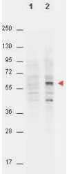
- Western Blot of Mouse anti-AKT pT308 antibody. Lane 1: non-phosphorylated AKT in untreated NIH/3T3 cells. Lane 2: phosphorylated AKT on PDGF stimulated NIH/3T3 cell lysates. Load: 15 µg per lane. Primary antibody: AKT pT308 antibody at a 1:4,000 dilution in TBS with 3% BSA, for 3 h at 4° C. Secondary antibody: peroxidase conjugated Gt-a-Mouse IgG (Fc) (p/n 610-1303) was used at a 1:40,000 dilution for 1 h at 4° C. Block: 3% BSA (p/n BSA-30) in TBS for 30 min at RT. Predicted/Observed size: (indicated by arrowhead at ~56 kDa). Other band(s): unspecific.
- Submitted by
- Invitrogen Antibodies (provider)
- Main image

- Experimental details
- Western Blot of Mouse anti-AKT pT308 antibody. Lane 1: non-phosphorylated AKT in untreated NIH/3T3 cells. Lane 2: phosphorylated AKT on PDGF stimulated NIH/3T3 cell lysates. Load: 15 µg per lane. Primary antibody: AKT pT308 antibody at a 1:4,000 dilution in TBS with 3% BSA, for 3 h at 4° C. Secondary antibody: peroxidase conjugated Gt-a-Mouse IgG (Fc) (p/n 610-1303) was used at a 1:40,000 dilution for 1 h at 4° C. Block: 3% BSA (p/n BSA-30) in TBS for 30 min at RT. Predicted/Observed size: (indicated by arrowhead at ~56 kDa). Other band(s): unspecific.

Supportive validation
- Submitted by
- Invitrogen Antibodies (provider)
- Main image
- Experimental details
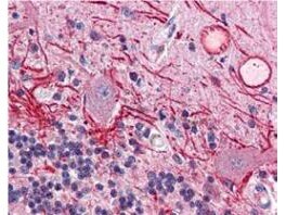
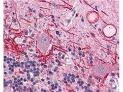
- Immunohistochemistry of Mouse anti-AKT pT308 antibody. Tissue: human brain cerebellum tissue (40X). Fixation: formalin fixed paraffin embedded. Antigen retrieval: not required. Primary antibody: AKT pT308antibody at 20 µg/mL for 1 h at RT. Secondary antibody: Peroxidase rabbit secondary antibody at 1:10,000 for 45 min at RT. Localization: staining of Purkinje neurons and cell processes in the cerebellum, cytosolic as well as occasionally nuclear. Staining: AKT pT308 as precipitated red signal with hematoxylin purple nuclear counterstain.