Explore
 Validate
 Learn
Learn Western blot
 ELISA
ELISAAntibody data
- Antibody Data
- Antigen structure
- References [5]
- Comments [0]
- Validations
- Western blot [3]
- Immunohistochemistry [1]
Submit
Validation data
Reference
Comment
Report error
- Product number
- NB600-608 - Provider product page

- Provider
- Novus Biologicals
- Proper citation
- Novus Cat#NB600-608, RRID:AB_10003539
- Product name
- Rabbit Polyclonal AKT1 Antibody
- Antibody type
- Polyclonal
- Description
- Unpurified.
- Reactivity
- Human
- Host
- Rabbit
- Vial size
- 0.1 ml
- Storage
- Store at -20C. Avoid freeze-thaw cycles.
Submitted references The centrosomal kinase NEK2 is a novel splicing factor kinase involved in cell survival.
Preferential expression of PAPPA in human preadipocytes from omental fat.
Low-intensity pulsed ultrasound activates the phosphatidylinositol 3 kinase/Akt pathway and stimulates the growth of chondrocytes in three-dimensional cultures: a basic science study.
Activation of the serine/threonine protein kinase Akt during the progression of Barrett neoplasia.
Silencing of caspase-8 and caspase-3 by RNA interference prevents vascular endothelial cell injury in mice with endotoxic shock.
Naro C, Barbagallo F, Chieffi P, Bourgeois CF, Paronetto MP, Sette C
Nucleic acids research 2014 Mar;42(5):3218-27
Nucleic acids research 2014 Mar;42(5):3218-27
Preferential expression of PAPPA in human preadipocytes from omental fat.
Davidge-Pitts C, Escande CJ, Conover CA
The Journal of endocrinology 2014 Jul;222(1):87-97
The Journal of endocrinology 2014 Jul;222(1):87-97
Low-intensity pulsed ultrasound activates the phosphatidylinositol 3 kinase/Akt pathway and stimulates the growth of chondrocytes in three-dimensional cultures: a basic science study.
Takeuchi R, Ryo A, Komitsu N, Mikuni-Takagaki Y, Fukui A, Takagi Y, Shiraishi T, Morishita S, Yamazaki Y, Kumagai K, Aoki I, Saito T
Arthritis research & therapy 2008;10(4):R77
Arthritis research & therapy 2008;10(4):R77
Activation of the serine/threonine protein kinase Akt during the progression of Barrett neoplasia.
Sagatys E, Garrett CR, Boulware D, Kelley S, Malafa M, Cheng JQ, Sebti S, Coppola D
Human pathology 2007 Oct;38(10):1526-31
Human pathology 2007 Oct;38(10):1526-31
Silencing of caspase-8 and caspase-3 by RNA interference prevents vascular endothelial cell injury in mice with endotoxic shock.
Matsuda N, Takano Y, Kageyama S, Hatakeyama N, Shakunaga K, Kitajima I, Yamazaki M, Hattori Y
Cardiovascular research 2007 Oct 1;76(1):132-40
Cardiovascular research 2007 Oct 1;76(1):132-40
No comments: Submit comment
Supportive validation
- Submitted by
- Novus Biologicals (provider)
- Main image

- Experimental details
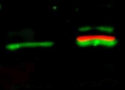
- Western Blot: AKT1 Antibody [NB600-608] - Lane 1: unstimulated NIH/3T3 lysates contain inactive unphosphorylated Akt1, green band. Lane 2: PDGF stimulated NIH/3T3 lysate contains both inactive (green band) and activated phosphorylated Akt1 (red band). 35 ug per lane. Rabbit anti-Akt (pan) and mouse anti-Akt pS473 specific antibodies at 1:1000 for overnight at 4C. Secondary antibody: DyLight 549 conjugated anti-rabbit IgG (green) and DyLight 649 conjugated anti-mouse IgG (red) secondary antibodies at 1:10,000 for 45 min at RT. Block: 5% BLOTTO overnight at 4C.
- Submitted by
- Novus Biologicals (provider)
- Main image
- Experimental details
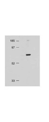
- Western Blot: AKT1 Antibody [NB600-608] - NIH/3T3 whole cell lysate, 20 ug. AKT1 antibody at 1:500 overnight at 4C. Secondary antibody: HRP conjugated Gt-a-Rabbit IgG at 1:10,000 preceded color development using Pierce Chemical's SuperSignal substrate. Block: MOPS buffer overnight at 4C. Predicted/Observed size: 56 kDa for AKT1.
- Submitted by
- Novus Biologicals (provider)
- Main image
- Experimental details
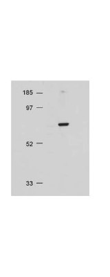
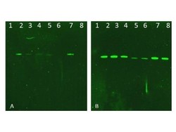
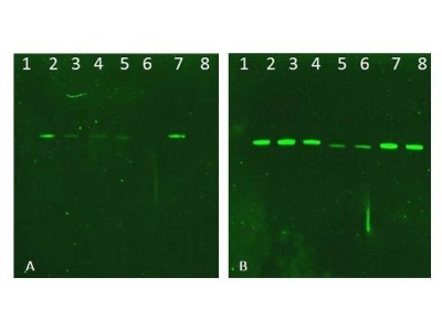
- Western Blot: AKT1 Antibody [NB600-608] - Lane 1: NIR MW protein ladder. Lane 2: AKT1, recombinant. Lane 3: AKT1, phosphatase-treated. Lane 4: AKT1, mutant T308A/S473A. Lane 5: AKT2, recombinant. Lane 6: AKT2, phosphatase-treated. Lane 7: AKT3, recombinant. Lane 8: AKT3, phosphatase-treated. Load: 50ng per lane. Blot A: Anti-Akt pT308 used at 1:2270, Blot B: Anti-Akt used 1:1000.
Supportive validation
- Submitted by
- Novus Biologicals (provider)
- Main image

- Experimental details
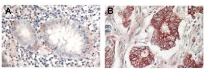
- Immunohistochemistry: AKT1 Antibody [NB600-608] - Analysis of FFPE Tissue: (A) normal colon tissue, (B) colon tumor tissue. Antigen retrieval not required. AKT1 antibody at 1:1,000 dilution for 1 h at RT. Peroxidase rabbit secondary antibody at 1:10,000 for 45 min at RT. AKT1 is nuclear. AKT1 as precipitated red signal with hematoxylin purple nuclear counterstain.