 Explore
 Validate
 Learn
Learn Western blot
 Immunocytochemistry
Antibody data
- Antibody Data
- Antigen structure
- References [2]
- Comments [0]
- Validations
- Immunocytochemistry [1]
- Immunoprecipitation [1]
- Immunohistochemistry [1]
- Other assay [6]
Submit
Validation data
Reference
Comment
Report error
- Product number
- PA5-29169 - Provider product page

- Provider
- Invitrogen Antibodies
- Product name
- AKT1 Polyclonal Antibody
- Antibody type
- Polyclonal
- Antigen
- Recombinant full-length protein
- Description
- Recommended positive controls: HA-AKT transfected HeLa cell, mouse liver. Predicted reactivity: Mouse (98%), Rat (97%), Zebrafish (84%), Xenopus laevis (90%), Cat (98%), Pig (96%), Chicken (96%), Sheep (96%), Rhesus Monkey (99%), Bovine (94%). Store product as a concentrated solution. Centrifuge briefly prior to opening the vial.
- Reactivity
- Human, Mouse
- Host
- Rabbit
- Isotype
- IgG
- Vial size
- 100 μL
- Concentration
- 0.47 mg/mL
- Storage
- Store at 4°C short term. For long term storage, store at -20°C, avoiding freeze/thaw cycles.
Submitted references Study on Toll-Like Receptor 2-Mediated Inflammation-Induced Familial Hypertension Combined with Hyperlipemia and Its Mechanism.
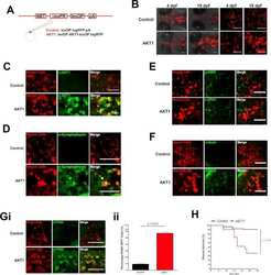
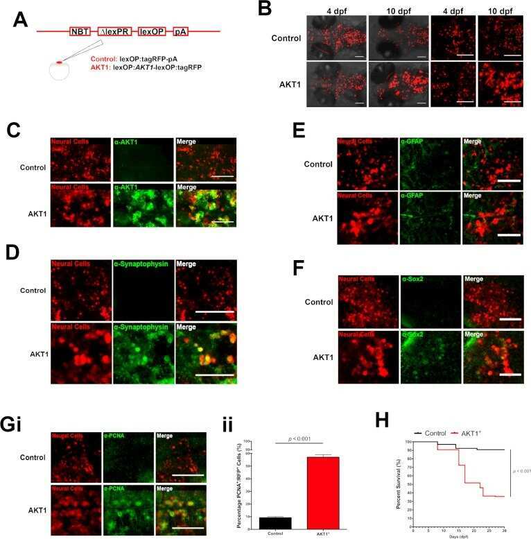
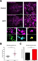
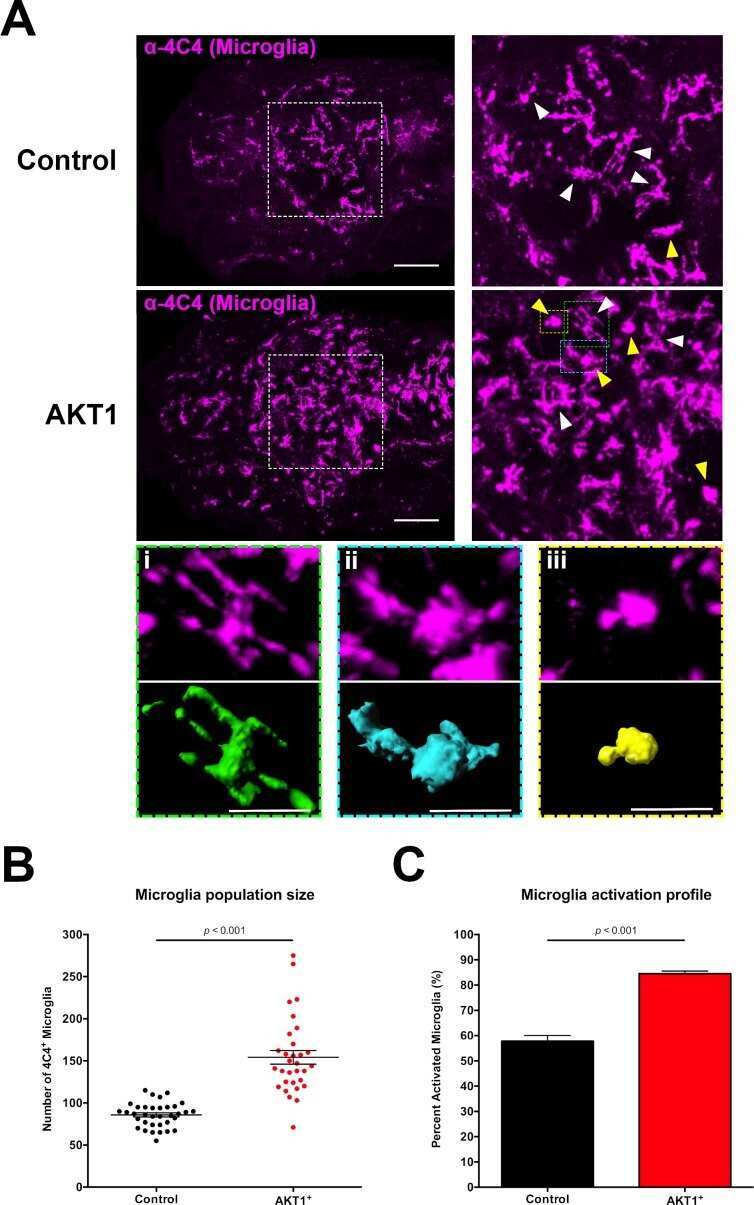
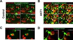
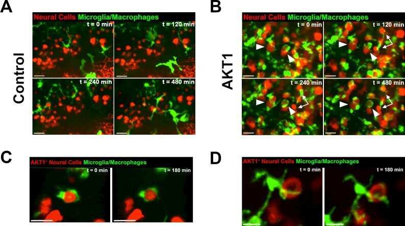
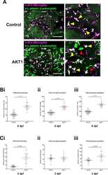
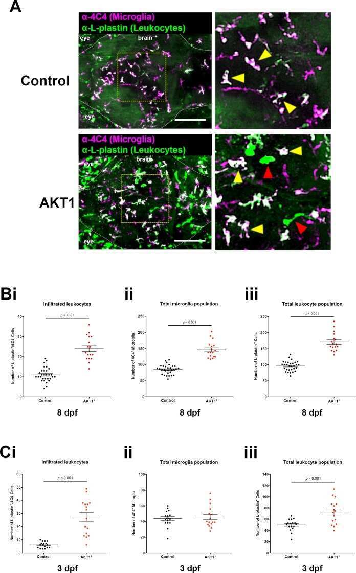
Tumor initiating cells induce Cxcr4-mediated infiltration of pro-tumoral macrophages into the brain.
Liu J, Li C, Wang Q, Hu H, Li C, Qian J
Journal of healthcare engineering 2022;2022:1473597
Journal of healthcare engineering 2022;2022:1473597
Tumor initiating cells induce Cxcr4-mediated infiltration of pro-tumoral macrophages into the brain.
Chia K, Mazzolini J, Mione M, Sieger D
eLife 2018 Feb 21;7
eLife 2018 Feb 21;7
No comments: Submit comment
Supportive validation
- Submitted by
- Invitrogen Antibodies (provider)
- Main image
- Experimental details
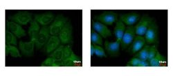
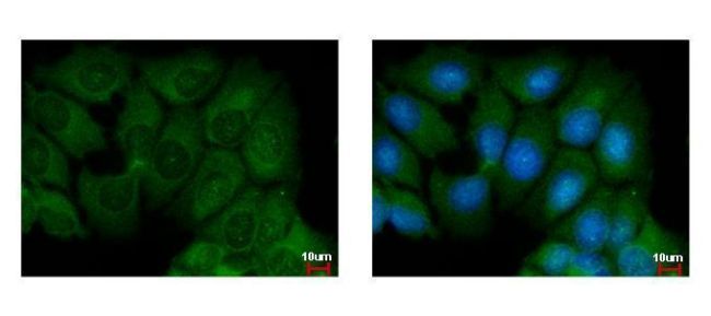
- Immunofluorescent analysis of AKT1 showing staining in the cytoplasm of MCF-7 cells. MCF-7 cells were fixed in ice-cold MeOH for 5 min and stained using an AKT1 polyclonal antibody (Product # PA5-29169) diluted at 1:500. Blue: Hoechst 33342 staining.
Supportive validation
- Submitted by
- Invitrogen Antibodies (provider)
- Main image
- Experimental details
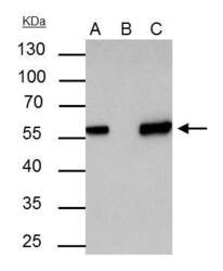
- Akt1 antibody immunoprecipitates Akt1 protein in IP experiments. IP samples: 30 µg whole cell extract of Akt1-transfected 293T cells. A. 30 µg whole cell extract of Akt1-protein expressing 293T cell. B. Control with 3 µg of preimmune Rabbit IgG. C. Immunoprecipitation of Akt1 protein by 3 µg Akt1 antibody (Product # PA5-29169). 10 % SDS-PAGE. The immunoprecipitated Akt1 protein was detected by Akt1 antibody (Product # PA5-29169) diluted at 1:5000.

Supportive validation
- Submitted by
- Invitrogen Antibodies (provider)
- Main image

- Experimental details
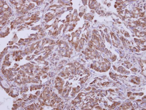
- Immunohistochemical analysis of paraffin-embedded human lung adenocarcinoma, using Akt1 (Product # PA5-29169) antibody at 1:250 dilution. Antigen Retrieval: EDTA based buffer, pH 8.0, 15 min.
Supportive validation
- Submitted by
- Invitrogen Antibodies (provider)
- Main image

- Experimental details
- Akt1 antibody immunoprecipitates Akt1 protein in IP experiments. IP samples: 30 µg whole cell extract of Akt1-transfected 293T cells. A. 30 µg whole cell extract of Akt1-protein expressing 293T cell. B. Control with 3 µg of preimmune Rabbit IgG. C. Immunoprecipitation of Akt1 protein by 3 µg Akt1 antibody (Product # PA5-29169). 10 % SDS-PAGE. The immunoprecipitated Akt1 protein was detected by Akt1 antibody (Product # PA5-29169) diluted at 1:5000.

- Submitted by
- Invitrogen Antibodies (provider)
- Main image
- Experimental details
- NULL
- Submitted by
- Invitrogen Antibodies (provider)
- Main image
- Experimental details
- NULL
- Submitted by
- Invitrogen Antibodies (provider)
- Main image
- Experimental details
- NULL
- Submitted by
- Invitrogen Antibodies (provider)
- Main image
- Experimental details
- NULL
- Submitted by
- Invitrogen Antibodies (provider)
- Main image

- Experimental details
- Akt1 antibody immunoprecipitates Akt1 protein in IP experiments. IP samples: 30 µg whole cell extract of Akt1-transfected 293T cells. A. 30 µg whole cell extract of Akt1-protein expressing 293T cell. B. Control with 3 µg of preimmune Rabbit IgG. C. Immunoprecipitation of Akt1 protein by 3 µg Akt1 antibody (Product # PA5-29169). 10 % SDS-PAGE. The immunoprecipitated Akt1 protein was detected by Akt1 antibody (Product # PA5-29169) diluted at 1:5000.
