Explore
 Validate
 Learn
Learn Western blot
Antibody data
- Antibody Data
- Antigen structure
- References [7]
- Comments [0]
- Validations
- Western blot [2]
- Immunocytochemistry [1]
- Immunohistochemistry [4]
Submit
Validation data
Reference
Comment
Report error
- Product number
- GTX28932 - Provider product page

- Provider
- GeneTex
- Proper citation
- GeneTex Cat#GTX28932, RRID:AB_367525
- Product name
- AKT (phospho Ser473) antibody
- Antibody type
- Polyclonal
- Reactivity
- Human, Mouse
- Host
- Rabbit
Submitted references Pterostilbene modulates the suppression of multidrug resistance protein 1 and triggers autophagic and apoptotic mechanisms in cisplatin-resistant human oral cancer CAR cells via AKT signaling.
AKT and its related molecular feature in aged mice skin.
Macrophages induce AKT/β-catenin-dependent Lgr5(+) stem cell activation and hair follicle regeneration through TNF.
Distinctively Expressed Cytokines by Three Different Inflammation Cells and Their Interaction with Keratinocytes in Wound Healing.
Prediabetes linked to excess glucagon in transgenic mice with pancreatic active AKT1.
Quercetin 3-O-methyl ether protects FL83B cells from copper induced oxidative stress through the PI3K/Akt and MAPK/Erk pathway.
The protective effects of spirulina in cyclophosphamide induced nephrotoxicity and urotoxicity in rats.
Chang HP, Lu CC, Chiang JH, Tsai FJ, Juan YN, Tsao JW, Chiu HY, Yang JS
International journal of oncology 2018 May;52(5):1504-1514
International journal of oncology 2018 May;52(5):1504-1514
AKT and its related molecular feature in aged mice skin.
Chen H, Wang X, Han J, Fan Z, Sadia S, Zhang R, Guo Y, Jiang Y, Wu Y
PloS one 2017;12(6):e0178969
PloS one 2017;12(6):e0178969
Macrophages induce AKT/β-catenin-dependent Lgr5(+) stem cell activation and hair follicle regeneration through TNF.
Wang X, Chen H, Tian R, Zhang Y, Drutskaya MS, Wang C, Ge J, Fan Z, Kong D, Wang X, Cai T, Zhou Y, Wang J, Wang J, Wang S, Qin Z, Jia H, Wu Y, Liu J, Nedospasov SA, Tredget EE, Lin M, Liu J, Jiang Y, Wu Y
Nature communications 2017 Mar 27;8:14091
Nature communications 2017 Mar 27;8:14091
Distinctively Expressed Cytokines by Three Different Inflammation Cells and Their Interaction with Keratinocytes in Wound Healing.
Wang J, Wang X, Chen H, Liu J, Tredget EE, Wu Y
Inflammation 2017 Dec;40(6):2151-2162
Inflammation 2017 Dec;40(6):2151-2162
Prediabetes linked to excess glucagon in transgenic mice with pancreatic active AKT1.
Albury-Warren TM, Pandey V, Spinel LP, Masternak MM, Altomare DA
The Journal of endocrinology 2016 Jan;228(1):49-59
The Journal of endocrinology 2016 Jan;228(1):49-59
Quercetin 3-O-methyl ether protects FL83B cells from copper induced oxidative stress through the PI3K/Akt and MAPK/Erk pathway.
Tseng HL, Li CJ, Huang LH, Chen CY, Tsai CH, Lin CN, Hsu HY
Toxicology and applied pharmacology 2012 Oct 1;264(1):104-13
Toxicology and applied pharmacology 2012 Oct 1;264(1):104-13
The protective effects of spirulina in cyclophosphamide induced nephrotoxicity and urotoxicity in rats.
Sinanoglu O, Yener AN, Ekici S, Midi A, Aksungar FB
Urology 2012 Dec;80(6):1392.e1-6
Urology 2012 Dec;80(6):1392.e1-6
No comments: Submit comment
Supportive validation
- Submitted by
- GeneTex (provider)
- Main image

- Experimental details
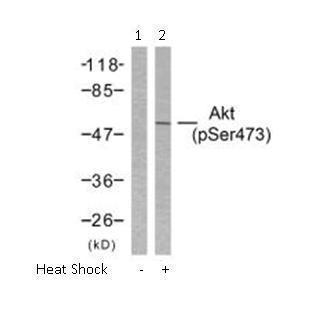
- Western blot analysis of extract from HeLa cells untreated or treated with heat shock using Akt (phospho-Ser473) antibody (GTX28932, Lane 1 and 2).
- Submitted by
- GeneTex (provider)
- Main image
- Experimental details
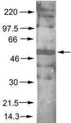
- Rabbit anti-AKT pS473 was used at a 1:200 dilution to detect phosphorylated AKT by Western blot. A nuclear extract from cells infected with adenovirus expressing nuclear-targeted AKT kinase was used.

Supportive validation
- Submitted by
- GeneTex (provider)
- Main image
- Experimental details
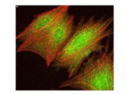
- Immunofluorescence Confocal Microscopy of Rabbit anti-AKT pS473 antibody (GTX28932). Tissue: cardiomyocytes infected with adenovirus expressing with wild-type AKT. Fixation: 0.5% PFA. Antigen retrieval: not required. Primary antibody: AKT pS473 antibody at 1:40 for 1 h at RT. Secondary antibody: texas-red conjugated rabbit secondary antibody at 1:10,000 for 45 min at RT. Localization: AKT pS473 is nuclear. Staining: AKT pS473 as green fluorescent signal with texas-red conjugated phalloidin (red) to label filamentous actin.

Supportive validation
- Submitted by
- GeneTex (provider)
- Main image
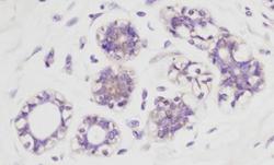
- Experimental details
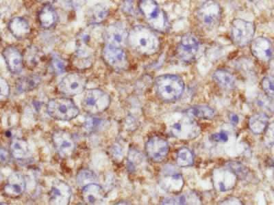
- Rabbit anti-AKT pS473 was used at a 1:100 dilution to detect AKT by immunohistochemistry in normal human breast tissue. AKT is weakly phosphorylated in normal tissue in the breast. The phosphorylated AKT is clearly localized in the cytoplasm. Rabbit anti-AKT pS473 antibody was used with no pretreatment of tissue. Tissue was formalin-fixed and paraffin embedded.
- Submitted by
- GeneTex (provider)
- Main image
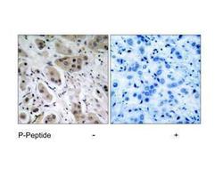
- Experimental details
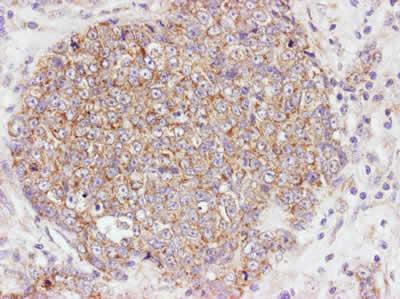
- Immunohistochemical analysis of paraffin- embedded human breast carcinoma tissue, using Akt (phospho-Ser473) antibody (GTX28932).
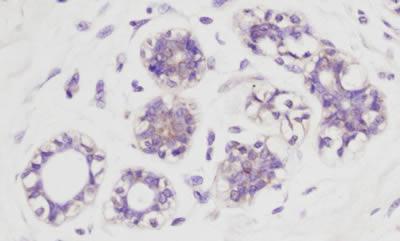
- Submitted by
- GeneTex (provider)
- Main image

- Experimental details
- Rabbit anti-AKT pS473 was used at a 1:100 dilution to detect AKT by immunohistochemistry in human breast tumor tissue. The staining is much stronger than the weak basal level of phosphorylation in normal breast.Rabbit anti-AKT pS473 antibody was used with no pretreatment of tissue.Tissue was formalin-fixed and paraffin embedded.
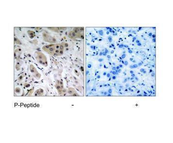
- Submitted by
- GeneTex (provider)
- Main image
- Experimental details
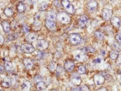
- This image is a magnification of the accompanying breast tumor image.