 Explore
 Validate
 Learn
Learn Western blot
Antibody data
- Antibody Data
- Antigen structure
- References [0]
- Comments [0]
- Validations
- Western blot [2]
- Immunohistochemistry [1]
Submit
Validation data
Reference
Comment
Report error
- Product number
- AF8375 - Provider product page

- Provider
- R&D Systems
- Product name
- Human NAC1 Antibody
- Antibody type
- Polyclonal
- Description
- Antigen Affinity-purified. Detects human NAC1 in direct ELISAs and Western Blots. In direct ELISAs, appoximately 100% cross-reactivity with recombinant mouse NAC1 is observed.
- Reactivity
- Human
- Host
- Sheep
- Conjugate
- Unconjugated
- Antigen sequence
Q96RE7- Isotype
- IgG
- Vial size
- 100 ug
- Storage
- Use a manual defrost freezer and avoid repeated freeze-thaw cycles. 12 months from date of receipt, -20 to -70 °C as supplied. 1 month, 2 to 8 °C under sterile conditions after reconstitution. 6 months, -20 to -70 °C under sterile conditions after reconstitution.
No comments: Submit comment
Supportive validation
- Submitted by
- R&D Systems (provider)
- Main image

- Experimental details
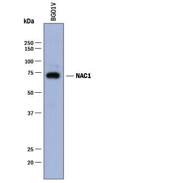
- Detection of Human NAC1 by Western Blot. Western blot shows lysates of BG01V human embryonic stem cells. PVDF membrane was probed with 1 µg/mL of Sheep Anti-Human NAC1 Antigen Affinity-purified Polyclonal Antibody (Catalog # AF8375) followed by HRP-conjugated Anti-Sheep IgG Secondary Antibody (Catalog # HAF016). A specific band was detected for NAC1 at approximately 72 kDa (as indicated). This experiment was conducted under reducing conditions and using Immunoblot Buffer Group 1.
- Submitted by
- R&D Systems (provider)
- Main image
- Experimental details
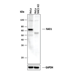
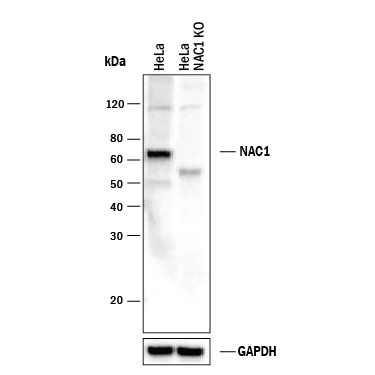
- Western Blot Shows Human NAC1 Specificity by Using Knockout Cell Line. Western blot shows lysates of HeLa human cervical epithelial carcinoma parental cell line and NAC1 knockout HeLa cell line (KO). PVDF membrane was probed with 1 µg/mL of Sheep Anti-Human NAC1 Antigen Affinity-purified Polyclonal Antibody (Catalog # AF8375) followed by HRP-conjugated Anti-Sheep IgG Secondary Antibody (Catalog # HAF016). A specific band was detected for NAC1 at approximately 68 kDa (as indicated) in the parental HeLa cell line, but is not detectable in knockout HeLa cell line. GAPDH (Catalog # AF5718) is shown as a loading control. This experiment was conducted under reducing conditions and using Immunoblot Buffer Group 1.
Supportive validation
- Submitted by
- R&D Systems (provider)
- Main image
- Experimental details
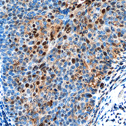
- NAC1 in Human Cervical Cancer Tissue. NAC1 was detected in immersion fixed paraffin-embedded sections of human cervical cancer tissue using Sheep Anti-Human NAC1 Antigen Affinity-purified Polyclonal Antibody (Catalog # AF8375) at 1 µg/mL overnight at 4 °C. Tissue was stained using the Anti-Sheep HRP-DAB Cell & Tissue Staining Kit (brown; Catalog # CTS019) and counterstained with hematoxylin (blue). Specific staining was localized to nuclei in cancer cells. View our protocol for Chromogenic IHC Staining of Paraffin-embedded Tissue Sections.
