Explore
 Validate
 Learn
Learn Western blot
 Immunohistochemistry
Antibody data
- Antibody Data
- Antigen structure
- References [1]
- Comments [0]
- Validations
- Western blot [2]
- Immunocytochemistry [3]
Submit
Validation data
Reference
Comment
Report error
- Product number
- PA5-34676 - Provider product page

- Provider
- Invitrogen Antibodies
- Product name
- ACAT1 Polyclonal Antibody
- Antibody type
- Polyclonal
- Antigen
- Recombinant protein fragment
- Description
- Recommended positive controls: 293T, A431, Molt-4, mouse brain, F31, F90. Predicted reactivity: Mouse (84%), Rat (83%), Zebrafish (80%), Rhesus Monkey (98%), Bovine (95%). Store product as a concentrated solution. Centrifuge briefly prior to opening the vial.
- Reactivity
- Human, Mouse
- Host
- Rabbit
- Isotype
- IgG
- Vial size
- 100 µL
- Concentration
- 1 mg/mL
- Storage
- Store at 4°C short term. For long term storage, store at -20°C, avoiding freeze/thaw cycles.
Submitted references Defining decreased protein succinylation of failing human cardiac myofibrils in ischemic cardiomyopathy.
Ali HR, Michel CR, Lin YH, McKinsey TA, Jeong MY, Ambardekar AV, Cleveland JC, Reisdorph R, Reisdorph N, Woulfe KC, Fritz KS
Journal of molecular and cellular cardiology 2020 Jan;138:304-317
Journal of molecular and cellular cardiology 2020 Jan;138:304-317
No comments: Submit comment
Supportive validation
- Submitted by
- Invitrogen Antibodies (provider)
- Main image
- Experimental details
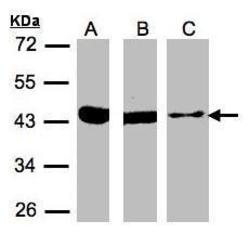
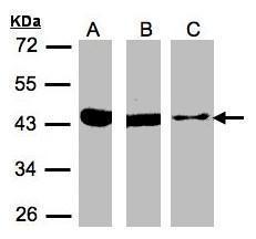
- Western Blot using ACAT1 Polyclonal Antibody (Product # PA5-34676). Sample (30 µg of whole cell lysate). A: 293T. B: A431. C: MOLT4SDS PAGE. Z. 10% SDS PAGE. ACAT1 Polyclonal Antibody (Product # PA5-34676) diluted at 1:500.
- Submitted by
- Invitrogen Antibodies (provider)
- Main image
- Experimental details
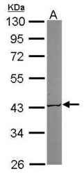
- Western Blot using ACAT1 Polyclonal Antibody (Product # PA5-34676). Sample (50 µg of whole cell lysate). Lane A: Mouse brain. 10% SDS PAGE. ACAT1 Polyclonal Antibody (Product # PA5-34676) diluted at 1:1,000.

Supportive validation
- Submitted by
- Invitrogen Antibodies (provider)
- Main image

- Experimental details
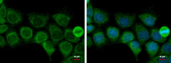
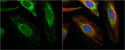
- Immunocytochemistry-Immunofluorescence analysis of ACAT1 was performed in HeLa cells fixed in 4% paraformaldehyde at RT for 15 min. Green: ACAT1 Polyclonal Antibody (Product # PA5-34676) diluted at 1:500. Red: alpha Tubulin, a cytoskeleton marker. Blue: Hoechst 33342 staining.

- Submitted by
- Invitrogen Antibodies (provider)
- Main image
- Experimental details
- Immunocytochemistry-Immunofluorescence analysis of ACAT1 was performed in HeLa cells fixed in 4% paraformaldehyde at RT for 15 min. Green: ACAT1 Polyclonal Antibody (Product # PA5-34676) diluted at 1:500. Red: alpha Tubulin, a cytoskeleton marker. Blue: Hoechst 33342 staining.

- Submitted by
- Invitrogen Antibodies (provider)
- Main image

- Experimental details
- ACAT1 Polyclonal Antibody detects ACAT1 protein at mitochondria by immunofluorescent analysis. Sample: A431 cells were fixed in 2% paraformaldehyde/culture medium at 37ºC for 30 min. Green: ACAT1 protein stained by ACAT1 Polyclonal Antibody (Product # PA5-34676) diluted at 1:500. Blue: Hoechst 33342 staining.