Explore
 Validate
 Learn
Learn Western blot
 Immunocytochemistry
Antibody data
- Antibody Data
- Antigen structure
- References [5]
- Comments [0]
- Validations
- Western blot [4]
- Immunohistochemistry [6]
Submit
Validation data
Reference
Comment
Report error
- Product number
- NBP1-89284 - Provider product page

- Provider
- Novus Biologicals
- Proper citation
- Novus Cat#NBP1-89284, RRID:AB_11054085
- Product name
- Rabbit Polyclonal ACAT1 Antibody
- Antibody type
- Polyclonal
- Description
- Immunogen affinity purified. Specificity of human ACAT1 antibody verified on a Protein Array containing target protein plus 383 other non-specific proteins.
- Reactivity
- Human, Porcine
- Host
- Rabbit
- Isotype
- IgG
- Vial size
- 0.1 ml
- Storage
- Store at 4C short term. Aliquot and store at -20C long term. Avoid freeze-thaw cycles.
Submitted references Antibodies biotinylated using a synthetic Z-domain from protein A provide stringent in situ protein detection.
Ketolytic and glycolytic enzymatic expression profiles in malignant gliomas: implication for ketogenic diet therapy.
Ketone body utilization drives tumor growth and metastasis.
Ketone bodies and two-compartment tumor metabolism: stromal ketone production fuels mitochondrial biogenesis in epithelial cancer cells.
Systematic validation of antibody binding and protein subcellular localization using siRNA and confocal microscopy.
Andersson S, Konrad A, Ashok N, Pontén F, Hober S, Asplund A
The journal of histochemistry and cytochemistry : official journal of the Histochemistry Society 2013 Nov;61(11):773-84
The journal of histochemistry and cytochemistry : official journal of the Histochemistry Society 2013 Nov;61(11):773-84
Ketolytic and glycolytic enzymatic expression profiles in malignant gliomas: implication for ketogenic diet therapy.
Chang HT, Olson LK, Schwartz KA
Nutrition & metabolism 2013 Jul 5;10(1):47
Nutrition & metabolism 2013 Jul 5;10(1):47
Ketone body utilization drives tumor growth and metastasis.
Martinez-Outschoorn UE, Lin Z, Whitaker-Menezes D, Howell A, Sotgia F, Lisanti MP
Cell cycle (Georgetown, Tex.) 2012 Nov 1;11(21):3964-71
Cell cycle (Georgetown, Tex.) 2012 Nov 1;11(21):3964-71
Ketone bodies and two-compartment tumor metabolism: stromal ketone production fuels mitochondrial biogenesis in epithelial cancer cells.
Martinez-Outschoorn UE, Lin Z, Whitaker-Menezes D, Howell A, Lisanti MP, Sotgia F
Cell cycle (Georgetown, Tex.) 2012 Nov 1;11(21):3956-63
Cell cycle (Georgetown, Tex.) 2012 Nov 1;11(21):3956-63
Systematic validation of antibody binding and protein subcellular localization using siRNA and confocal microscopy.
Stadler C, Hjelmare M, Neumann B, Jonasson K, Pepperkok R, Uhlén M, Lundberg E
Journal of proteomics 2012 Apr 3;75(7):2236-51
Journal of proteomics 2012 Apr 3;75(7):2236-51
No comments: Submit comment
Supportive validation
- Submitted by
- Novus Biologicals (provider)
- Main image

- Experimental details
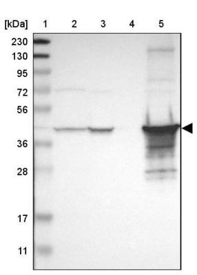
- Western Blot: ACAT1 Antibody [NBP1-89284] - Lane 1: Marker [kDa] 230, 130, 95, 72, 56, 36, 28, 17, 11. Lane 2: Human cell line RT-4. Lane 3: Human cell line U-251MG sp. Lane 4: Human plasma (IgG/HSA depleted). Lane 5: Human liver tissue
- Submitted by
- Novus Biologicals (provider)
- Main image

- Experimental details
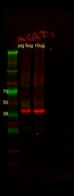
- Western Blot: ACAT1 Antibody [NBP1-89284] - ACAT1 porcine cerebral cortex. Image from verified customer review.
- Submitted by
- Novus Biologicals (provider)
- Main image
- Experimental details
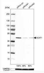
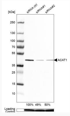
- Western Blot: ACAT1 Antibody [NBP1-89284] - Analysis in Caco-2 cells transfected with control siRNA, target specific siRNA probe #1 and #2, using Anti-ACAT1 antibody. Remaining relative intensity is presented. Loading control: Anti-PPIB.
- Submitted by
- Novus Biologicals (provider)
- Main image
- Experimental details
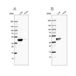
- Western Blot: ACAT1 Antibody [NBP1-89284] - Analysis using Anti-ACAT1 antibody NBP1-89284 (A) shows similar pattern to independent antibody NBP1-89285 (B).

Supportive validation
- Submitted by
- Novus Biologicals (provider)
- Main image

- Experimental details
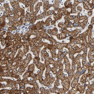
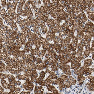
- Immunohistochemistry-Paraffin: ACAT1 Antibody [NBP1-89284] - Staining of human liver shows strong cytoplasmic positivity in hepatocytes.
- Submitted by
- Novus Biologicals (provider)
- Main image
- Experimental details
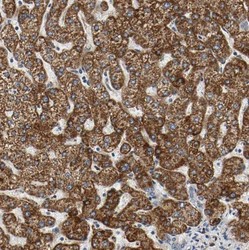
- Immunohistochemistry-Paraffin: ACAT1 Antibody [NBP1-89284] - Staining of human liver shows strong positivity in mitochondria in hepatocytes.
- Submitted by
- Novus Biologicals (provider)
- Main image
- Experimental details
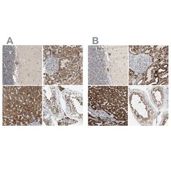
- Immunohistochemistry-Paraffin: ACAT1 Antibody [NBP1-89284] - Staining of human cerebellum, kidney, liver and testis using Anti-ACAT1 antibody NBP1-89284 (A) shows similar protein distribution across tissues to independent antibody NBP1-89285 (B).

- Submitted by
- Novus Biologicals (provider)
- Main image
- Experimental details
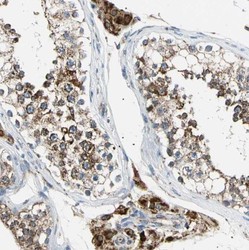
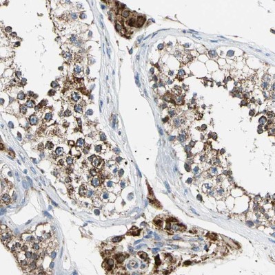
- Immunohistochemistry-Paraffin: ACAT1 Antibody [NBP1-89284] - Staining of human testis shows strong positivity in mitochondria in cells in seminiferous ducts.
- Submitted by
- Novus Biologicals (provider)
- Main image
- Experimental details
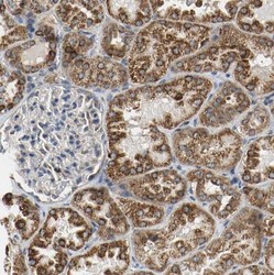
- Immunohistochemistry-Paraffin: ACAT1 Antibody [NBP1-89284] - Staining of human kidney shows strong positivity in mitochondria in cells in tubules.

- Submitted by
- Novus Biologicals (provider)
- Main image
- Experimental details
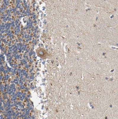
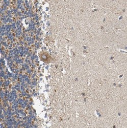
- Immunohistochemistry-Paraffin: ACAT1 Antibody [NBP1-89284] - Staining of human cerebellum shows moderate positivity in mitochondria in Purkinje cells.