Explore
 Validate
 Learn
Learn Western blot
Antibody data
- Antibody Data
- Antigen structure
- References [0]
- Comments [0]
- Validations
- Western blot [4]
- Immunocytochemistry [1]
Submit
Validation data
Reference
Comment
Report error
- Product number
- PA5-28479 - Provider product page

- Provider
- Invitrogen Antibodies
- Product name
- Staufen Polyclonal Antibody
- Antibody type
- Polyclonal
- Antigen
- Recombinant protein fragment
- Description
- Recommended positive controls: 293T, A431, H1299, HepG2, Molt-4, Raji, Neuro2A, 293 (input), Stau1-IP (GTX106566), Post-IP lysate from control rabbit IgG-IP, Post-IP lysate from Stau1-IP.
- Concentration
- 0.93 mg/mL
No comments: Submit comment
Supportive validation
- Submitted by
- Invitrogen Antibodies (provider)
- Main image

- Experimental details
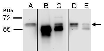
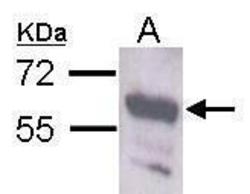
- Western blot analysis of Staufen using A) 293 (input) (B) control rabbit IgG-IP (C) Stau1-IP (D) Post-IP lysate from control rabbit IgG-IP and E) Post-IP lysate from Stau1-IP. Samples were loaded onto a 7.5% SDS-PAGE gel and probed with a Staufen polyclonal antibody (Product # PA5-28479) at a dilution of 1:3000.
- Submitted by
- Invitrogen Antibodies (provider)
- Main image
- Experimental details
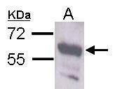
- Western blot analysis of Staufen using 30 µg of HepG2 lysate. Samples were loaded onto a 7.5% SDS-PAGE gel and probed with a Staufen polyclonal antibody (Product # PA5-28479) at a dilution of 1:5000.
- Submitted by
- Invitrogen Antibodies (provider)
- Main image
- Experimental details
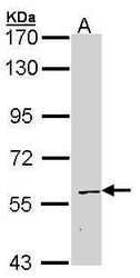
- Western Blot using Staufen Polyclonal Antibody (Product # PA5-28479). Sample (30 µg of whole cell lysate). Lane A: Neuro2A. Staufen Polyclonal Antibody (Product # PA5-28479) diluted at 1:1,000. The HRP-conjugated anti-rabbit IgG antibody was used to detect the primary antibody.
- Submitted by
- Invitrogen Antibodies (provider)
- Main image
- Experimental details
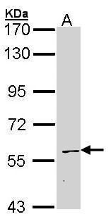
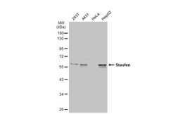
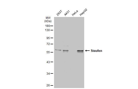
- Western Blot using Staufen Polyclonal Antibody (Product # PA5-28479). Various whole cell extracts (30 µg) were separated by 10% SDS-PAGE, and the membrane was blotted with Staufen Polyclonal Antibody (Product # PA5-28479) diluted at 1:5,000. The HRP-conjugated anti-rabbit IgG antibody was used to detect the primary antibody.
Supportive validation
- Submitted by
- Invitrogen Antibodies (provider)
- Main image
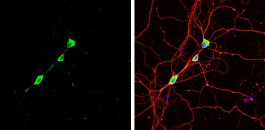
- Experimental details
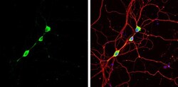
- Immunocytochemistry-Immunofluorescence analysis of Staufen was performed in DIV9 rat E18 primary hippocampal neuron cells fixed in 4% paraformaldehyde at RT for 15 min. Green: Staufen Polyclonal Antibody (Product # PA5-28479) diluted at 1:500. Red: beta Tubulin 3/ Tuj1, stained by beta Tubulin 3/ Tuj1 antibody. Blue: Fluoroshield with DAPI.