 Explore
 Validate
 Learn
Learn Western blot
 Immunohistochemistry
Antibody data
- Antibody Data
- Antigen structure
- References [17]
- Comments [0]
- Validations
- Immunohistochemistry [1]
Submit
Validation data
Reference
Comment
Report error
- Product number
- HPA006641 - Provider product page

- Provider
- Atlas Antibodies
- Proper citation
- Atlas Antibodies Cat#HPA006641, RRID:AB_1079874
- Product name
- Anti-SCGN
- Antibody type
- Polyclonal
- Description
- Polyclonal Antibody against Human SCGN, Gene description: secretagogin, EF-hand calcium binding protein, Alternative Gene Names: CALBL, DJ501N12.8, SECRET, SEGN, Validated applications: WB, IHC, Uniprot ID: O76038, Storage: Store at +4°C for short term storage. Long time storage is recommended at -20°C.
- Reactivity
- Human, Mouse
- Host
- Rabbit
- Conjugate
- Unconjugated
- Isotype
- IgG
- Vial size
- 100 µl
- Concentration
- 0.2 mg/ml
- Storage
- Store at +4°C for short term storage. Long time storage is recommended at -20°C.
- Handling
- The antibody solution should be gently mixed before use.
Submitted references Meningioma classification by immunohistochemistry: A replicability study.
Postnatal Developmental Expression Profile Classifies the Indusium Griseum as a Distinct Subfield of the Hippocampal Formation
A clinically applicable integrative molecular classification of meningiomas
RNA-Seq Analysis Reveals an Essential Role of the Tyrosine Metabolic Pathway and Inflammation in Myopia-Induced Retinal Degeneration in Guinea Pigs
Ferret brain possesses young interneuron collections equivalent to human postnatal migratory streams
Immature excitatory neurons develop during adolescence in the human amygdala
Secretagogin is increased in plasma from type 2 diabetes patients and potentially reflects stress and islet dysfunction
Deafferented Adult Rod Bipolar Cells Create New Synapses with Photoreceptors to Restore Vision
Calcium Binding and Disulfide Bonds Regulate the Stability of Secretagogin towards Thermal and Urea Denaturation
Neuronal calcium-binding proteins 1/2 localize to dorsal root ganglia and excitatory spinal neurons and are regulated by nerve injury
Distribution of secretagogin-containing neurons in the basal forebrain of mice, with special reference to the cholinergic corticopetal system
Novel pancreatic beta cell-specific proteins: Antibody-based proteomics for identification of new biomarker candidates
The renaissance of Ca2+-binding proteins in the nervous system: secretagogin takes center stage
Clusters of secretagogin-expressing neurons in the aged human olfactory tract lack terminal differentiation
Secretagogin is a Ca2+‐binding protein identifying prospective extended amygdala neurons in the developing mammalian telencephalon
Secretagogin is a Ca 2+ -binding protein specifying subpopulations of telencephalic neurons
Tissue profiling of the mammalian central nervous system using human antibody-based proteomics.
Näslund O, Lipatnikova A, Dénes A, Lindskog C, Bontell TO, Smits A, Jakola AS, Corell A
Brain & spine 2023;3:101711
Brain & spine 2023;3:101711
Postnatal Developmental Expression Profile Classifies the Indusium Griseum as a Distinct Subfield of the Hippocampal Formation
Sanders M, Petrasch-Parwez E, Habbes H, Düring M, Förster E
Frontiers in Cell and Developmental Biology 2021;8
Frontiers in Cell and Developmental Biology 2021;8
A clinically applicable integrative molecular classification of meningiomas
Nassiri F, Liu J, Patil V, Mamatjan Y, Wang J, Hugh-White R, Macklin A, Khan S, Singh O, Karimi S, Corona R, Liu L, Chen C, Chakravarthy A, Wei Q, Mehani B, Suppiah S, Gao A, Workewych A, Tabatabai G, Boutros P, Bader G, de Carvalho D, Kislinger T, Aldape K, Zadeh G
Nature 2021;597(7874):119-125
Nature 2021;597(7874):119-125
RNA-Seq Analysis Reveals an Essential Role of the Tyrosine Metabolic Pathway and Inflammation in Myopia-Induced Retinal Degeneration in Guinea Pigs
Zeng L, Li X, Liu J, Liu H, Xu H, Yang Z
International Journal of Molecular Sciences 2021;22(22):12598
International Journal of Molecular Sciences 2021;22(22):12598
Ferret brain possesses young interneuron collections equivalent to human postnatal migratory streams
Ellis J, Sorrells S, Mikhailova S, Chavali M, Chang S, Sabeur K, Mcquillen P, Rowitch D
Journal of Comparative Neurology 2019;527(17):2843-2859
Journal of Comparative Neurology 2019;527(17):2843-2859
Immature excitatory neurons develop during adolescence in the human amygdala
Sorrells S, Paredes M, Velmeshev D, Herranz-Pérez V, Sandoval K, Mayer S, Chang E, Insausti R, Kriegstein A, Rubenstein J, Manuel Garcia-Verdugo J, Huang E, Alvarez-Buylla A
Nature Communications 2019;10(1)
Nature Communications 2019;10(1)
Secretagogin is increased in plasma from type 2 diabetes patients and potentially reflects stress and islet dysfunction
Fiorina P, Hansson S, Zhou A, Vachet P, Eriksson J, Pereira M, Skrtic S, Jongsma Wallin H, Ericsson-Dahlstrand A, Karlsson D, Ahnmark A, Sörhede Winzell M, Magnone M, Davidsson P
PLOS ONE 2018;13(4):e0196601
PLOS ONE 2018;13(4):e0196601
Deafferented Adult Rod Bipolar Cells Create New Synapses with Photoreceptors to Restore Vision
Beier C, Hovhannisyan A, Weiser S, Kung J, Lee S, Lee D, Huie P, Dalal R, Palanker D, Sher A
The Journal of Neuroscience 2017;37(17):4635-4644
The Journal of Neuroscience 2017;37(17):4635-4644
Calcium Binding and Disulfide Bonds Regulate the Stability of Secretagogin towards Thermal and Urea Denaturation
Garriga P, Sanagavarapu K, Weiffert T, Ní Mhurchú N, O’Connell D, Linse S
PLOS ONE 2016;11(11):e0165709
PLOS ONE 2016;11(11):e0165709
Neuronal calcium-binding proteins 1/2 localize to dorsal root ganglia and excitatory spinal neurons and are regulated by nerve injury
Zhang M, Tortoriello G, Hsueh B, Tomer R, Ye L, Mitsios N, Borgius L, Grant G, Kiehn O, Watanabe M, Uhlén M, Mulder J, Deisseroth K, Harkany T, Hökfelt T
Proceedings of the National Academy of Sciences 2014;111(12)
Proceedings of the National Academy of Sciences 2014;111(12)
Distribution of secretagogin-containing neurons in the basal forebrain of mice, with special reference to the cholinergic corticopetal system
Gyengesi E, Andrews Z, Paxinos G, Zaborszky L
Brain Research Bulletin 2013;94
Brain Research Bulletin 2013;94
Novel pancreatic beta cell-specific proteins: Antibody-based proteomics for identification of new biomarker candidates
Lindskog C, Korsgren O, Pontén F, Eriksson J, Johansson L, Danielsson A
Journal of Proteomics 2012;75(9):2611-2620
Journal of Proteomics 2012;75(9):2611-2620
The renaissance of Ca2+-binding proteins in the nervous system: secretagogin takes center stage
Alpár A, Attems J, Mulder J, Hökfelt T, Harkany T
Cellular Signalling 2012;24(2):378-387
Cellular Signalling 2012;24(2):378-387
Clusters of secretagogin-expressing neurons in the aged human olfactory tract lack terminal differentiation
Attems J, Alpar A, Spence L, McParland S, Heikenwalder M, Uhlén M, Tanila H, Hökfelt T, Harkany T
Proceedings of the National Academy of Sciences 2012;109(16):6259-6264
Proceedings of the National Academy of Sciences 2012;109(16):6259-6264
Secretagogin is a Ca2+‐binding protein identifying prospective extended amygdala neurons in the developing mammalian telencephalon
Mulder J, Spence L, Tortoriello G, DiNieri J, Uhlén M, Shui B, Kotlikoff M, Yanagawa Y, Aujard F, Hökfelt T, Hurd Y, Harkany T
European Journal of Neuroscience 2010;31(12):2166-2177
European Journal of Neuroscience 2010;31(12):2166-2177
Secretagogin is a Ca 2+ -binding protein specifying subpopulations of telencephalic neurons
Mulder J, Zilberter M, Spence L, Tortoriello G, Uhlén M, Yanagawa Y, Aujard F, Hökfelt T, Harkany T
Proceedings of the National Academy of Sciences 2009;106(52):22492-22497
Proceedings of the National Academy of Sciences 2009;106(52):22492-22497
Tissue profiling of the mammalian central nervous system using human antibody-based proteomics.
Mulder J, Björling E, Jonasson K, Wernérus H, Hober S, Hökfelt T, Uhlén M
Molecular & cellular proteomics : MCP 2009 Jul;8(7):1612-22
Molecular & cellular proteomics : MCP 2009 Jul;8(7):1612-22
No comments: Submit comment
Supportive validation
- Submitted by
- Atlas Antibodies (provider)
- Enhanced method
- Orthogonal validation
- Main image
- Experimental details
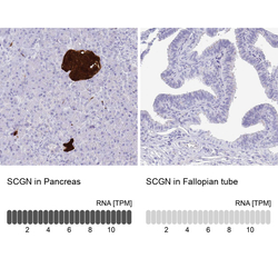
- Immunohistochemistry analysis in human pancreas and fallopian tube tissues using HPA006641 antibody. Corresponding SCGN RNA-seq data are presented for the same tissues.
- Sample type
- Human
- Protocol
- Protocol
