Explore
 Validate
 Learn
Learn Western blot
 Immunocytochemistry
Antibody data
- Antibody Data
- Antigen structure
- References [1]
- Comments [0]
- Validations
- Immunocytochemistry [1]
- Other assay [1]
Submit
Validation data
Reference
Comment
Report error
- Product number
- PA5-37361 - Provider product page

- Provider
- Invitrogen Antibodies
- Product name
- DDB2 Polyclonal Antibody
- Antibody type
- Polyclonal
- Antigen
- Recombinant full-length protein
- Description
- This antibody detects endogenous protein at a molecular weight of 48 kDa. Purity is >95% by SDS-PAGE.
- Reactivity
- Human, Mouse, Rat
- Host
- Rabbit
- Isotype
- IgG
- Vial size
- 100 μL
- Concentration
- 1 mg/mL
- Storage
- Store at 4°C short term. For long term storage, store at -20°C, avoiding freeze/thaw cycles.
Submitted references ROS-Induced Activation of DNA Damage Responses Drives Senescence-Like State in Postmitotic Cochlear Cells: Implication for Hearing Preservation.
Benkafadar N, François F, Affortit C, Casas F, Ceccato JC, Menardo J, Venail F, Malfroy-Camine B, Puel JL, Wang J
Molecular neurobiology 2019 Aug;56(8):5950-5969
Molecular neurobiology 2019 Aug;56(8):5950-5969
No comments: Submit comment
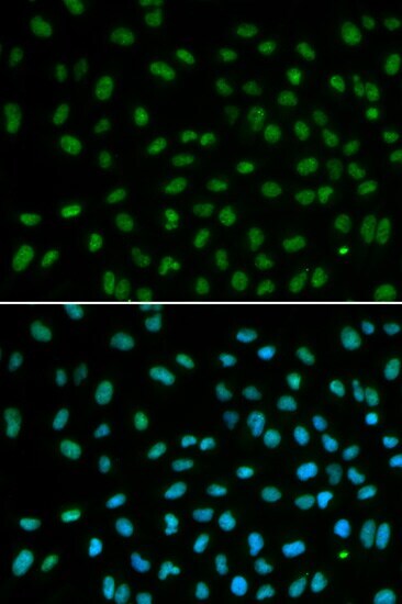
Supportive validation
- Submitted by
- Invitrogen Antibodies (provider)
- Main image
- Experimental details
- Immunofluorescence analysis of DDB2 in MCF7 cells. Samples were incubated with DDB2 polyclonal antibody (Product # PA5-37361).
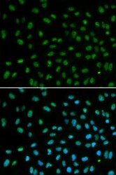
Supportive validation
- Submitted by
- Invitrogen Antibodies (provider)
- Main image
- Experimental details
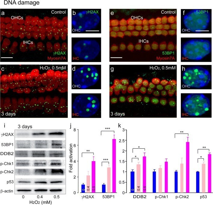
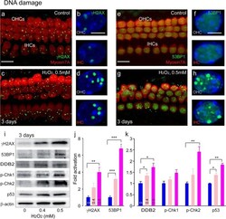
- Fig. 3 DNA damage and DNA damage responses upon H 2 O 2 challenge. a , c , e , g Confocal images showing the basal region of the organ of Corti cultures treated with either culture medium alone ( a , e ) or containing 0.5 mM H 2 O 2 ( c , g ) for 5 h before being maintained in culture medium alone for 3 days. The samples were then immunolabeled for myosin 7A (red, a , c , e , g ), gammaH2AX (green, a and c ) and 53BP1 (green, e and g ). Scale bars: a , c , e and g = 10 mum. b , d , f , h Higher magnification images of representative OHC and IHC nuclei from all conditions tested. Scale bar = 2.5 mum. i Representative Western blot analysis using antibodies against gammaH2AX, 53BP1, DDB2, p-Chk1, p-Chk2, p53, and beta-actin in whole cochlear extracts. j , k Histograms representing the levels of gammaH2AX, 53BP1, DDB2, p-Chk1, p-Chk2, and p53 in control and in 0.4 and 0.5 mM H 2 O 2 -exposed groups ( n = 6 cochleae per condition). beta-Actin served as a loading control. Data are expressed as mean +- SEM. One-way ANOVA test followed by post hoc Tukey's test (* P