Explore
 Validate
 Learn
Learn Western blot
 Immunocytochemistry
Antibody data
- Antibody Data
- Antigen structure
- References [1]
- Comments [0]
- Validations
- Immunocytochemistry [2]
- Flow cytometry [2]
- Other assay [2]
Submit
Validation data
Reference
Comment
Report error
- Product number
- PA5-79143 - Provider product page

- Provider
- Invitrogen Antibodies
- Product name
- DDB2 Polyclonal Antibody
- Antibody type
- Polyclonal
- Antigen
- Recombinant full-length protein
- Description
- Reconstitute with 0.2 mL of distilled water to yield a concentration of 500 µg/mL. Positive Control - WB: A431 whole cell, SW620 whole cell, HELA whole cell, JURKAT whole cell. ICC/IF: U20S cell. Flow: 293T cell.
- Reactivity
- Human
- Host
- Rabbit
- Isotype
- IgG
- Vial size
- 100 μg
- Concentration
- 500 μg/mL
- Storage
- -20°C
Submitted references DDB2 regulates DNA replication through PCNA-independent degradation of CDT2.
Wu X, Yu M, Zhang Z, Leng F, Ma Y, Xie N, Lu F
Cell & bioscience 2021 Feb 8;11(1):34
Cell & bioscience 2021 Feb 8;11(1):34
No comments: Submit comment
Supportive validation
- Submitted by
- Invitrogen Antibodies (provider)
- Main image

- Experimental details
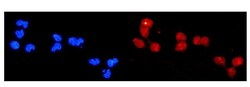
- Immunocytochemistry analysis of DDB2 using anti-DDB2 antibody (Product # PA5-79143). DDB2 was detected in a section of U2OS cells. Enzyme antigen retrieval was performed using IHC enzyme antigen retrieval reagent for 15 mins. The cells were blocked with 10% goat serum and then incubated with 2μg/mL rabbit anti-DDB2 antibody (Product # PA5-79143)overnight at 4°C. DyLight®550 Conjugated Goat Anti-Rabbit IgG was used as secondary antibody at 1:100 dilution and incubated for 30 minutes at 37°C. The section was counterstained with DAPI. Visualize using a fluorescence microscope and filter sets appropriate for the label used.

- Submitted by
- Invitrogen Antibodies (provider)
- Main image
- Experimental details
- Immunocytochemistry analysis of DDB2 using anti-DDB2 antibody (Product # PA5-79143). DDB2 was detected in a section of U2OS cells. Enzyme antigen retrieval was performed using IHC enzyme antigen retrieval reagent for 15 mins. The cells were blocked with 10% goat serum and then incubated with 2μg/mL rabbit anti-DDB2 antibody (Product # PA5-79143)overnight at 4°C. DyLight®550 Conjugated Goat Anti-Rabbit IgG was used as secondary antibody at 1:100 dilution and incubated for 30 minutes at 37°C. The section was counterstained with DAPI. Visualize using a fluorescence microscope and filter sets appropriate for the label used.

Supportive validation
- Submitted by
- Invitrogen Antibodies (provider)
- Main image
- Experimental details
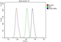
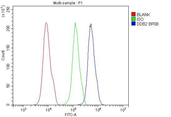
- Flow Cytometry of DDB2 in 293T cells (blue line), isotype control rabbit IgG (green line) and unlabeled (red line). Samples were blocked with 10% goat serum, incubated with DDB2 Polyclonal Antibody (Product # PA5-79143) at a dilution of 1 μg (per 1x10^6 cells), followed by DyLight®488 conjugated goat anti-rabbit IgG (for 30 minutes at 20°C) using 5-10 μg (per 1x10^6 cells) dilution.

- Submitted by
- Invitrogen Antibodies (provider)
- Main image

- Experimental details
- Flow Cytometry of DDB2 in 293T cells (blue line), isotype control rabbit IgG (green line) and unlabeled (red line). Samples were blocked with 10% goat serum, incubated with DDB2 Polyclonal Antibody (Product # PA5-79143) at a dilution of 1 μg (per 1x10^6 cells), followed by DyLight®488 conjugated goat anti-rabbit IgG (for 30 minutes at 20°C) using 5-10 μg (per 1x10^6 cells) dilution.
Supportive validation
- Submitted by
- Invitrogen Antibodies (provider)
- Main image
- Experimental details
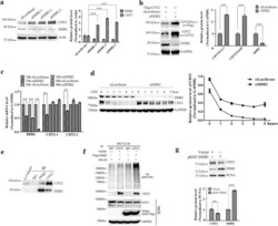
- Fig. 1 CRL DDB2 is a new E3 ubiquitin ligase of CDT2. a The protein level of CDT2 was accumulated when DDB2 was silenced. HCT116 cells were transfected with luciferase and DDB2 specific siRNAs for 48 h and subjected to Western blot. Actin was taken as loading control. Right panel: the relative protein levels of CDT2 and DDB2 were quantified by Gel-pro analyzer 4.0, and the P value was calculated by the two-side Student's t-test (*** indicated P < 0.001). The error bars denoted standard deviation (SD). b Silencing of DDB2 accumulated exogenous CDT2. HCT116 cells were treated with indicated siRNAs for 18 h, and then transfected with pCMV10-3Flag-CDT2 by polyetherimide and cultured for another 40 h. The protein levels of CDT2 and DDB2 were analyzed by Western blot. Exogenous CDT2 was detected by anti-Flag antibody. The relative protein level of CDT2 was measured using Gel-pro analyzer 4.0 and plotted on the right panel. *** indicated P < 0.001. The error bar indicated SD. c DDB2 had negligible effects on CDT2 transcription. HCT116 cells were transfected with indicated siRNAs, and mRNAs were extracted at 24 h, 36 h and 48 h respectively. The mRNA levels of interested genes were measured by RT-qPCR. Two pairs of specific primers targeting to CDT2 were used to quantify mRNA level of CDT2 . Student's t-test was used to calculate P value (** indicated P < 0.01, *** indicated P < 0.001). The error bars denoted SD. d Down regulation of DDB2 prolonged the half-life of CDT2. HCT116 cells

- Submitted by
- Invitrogen Antibodies (provider)
- Main image
- Experimental details
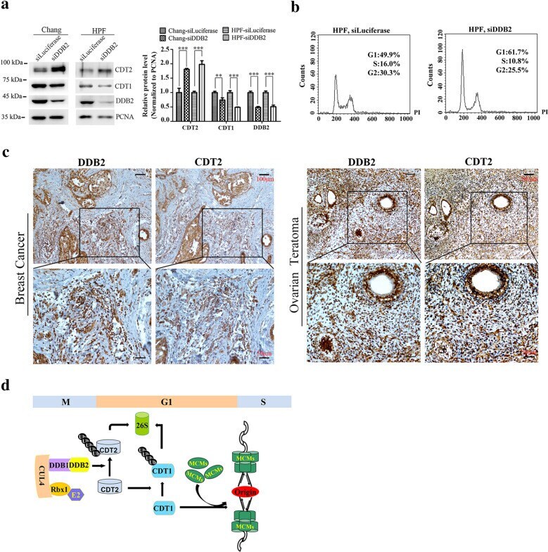
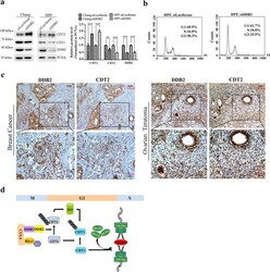
- Fig. 5 DDB2 mediated proteolysis of CDT2 may corelated with prognostic in cancer patients. a DDB2 promoted CDT2 proteolysis in Chang liver and CCC-HPF-1 cells. Chang liver and CCC-HPF-1 cells were treated with siRNAs of luciferase and DDB2 for 48 h, and harvested for Western blot. PCNA was taken as loading control. The relative protein levels of CDT2, CDT1 and DDB2 were normalized and plotted on the right panel. ** Denoted P < 0.01; *** denoted P < 0.001, and the error bars indicated SD. b CCC-HPF-1 cells were arrested in G1 phase after DDB2 silencing. FACS analysis of DNA contents in CCC-HPF-1 cells after treatment with siRNAs of luciferase and DDB2 for 48 h. c Expression of CDT2 and DDB2 in ovarian teratoma and breast cancer tissues detected by IHC. Scale bar: 50 um and 100 um. d The schematic model of CRL4 DDB2 ubiquitin ligase regulates DNA replication initiation through degrading CDT2. In late M and G1 phase, the degradation of CDT2 mediated by CRL4 DDB2 ubiquitin ligase stabilizes CDT1. Accumulation of CDT1 promotes recruitment of MCMs onto origins and assembly of pre-replication complex