Explore
 Validate
 Learn
Learn Western blot
 Immunohistochemistry
Antibody data
- Antibody Data
- Antigen structure
- References [1]
- Comments [0]
- Validations
- Immunohistochemistry [1]
- Other assay [2]
Submit
Validation data
Reference
Comment
Report error
- Product number
- 702595 - Provider product page

- Provider
- Invitrogen Antibodies
- Product name
- GPR68 Recombinant Rabbit Monoclonal Antibody (16H23L16)
- Antibody type
- Monoclonal
- Antigen
- Synthetic peptide
- Description
- This antibody is predicted to react with Monkey, Rat, Mouse, Horse. Recombinant rabbit monoclonal antibodies are produced using in vitro expression systems. The expression systems are developed by cloning in the specific antibody DNA sequences from immunoreactive rabbits. Then, individual clones are screened to select the best candidates for production. The advantages of using recombinant rabbit monoclonal antibodies include: better specificity and sensitivity, lot-to-lot consistency, animal origin-free formulations, and broader immunoreactivity to diverse targets due to larger rabbit immune repertoire.
- Reactivity
- Human
- Host
- Rabbit
- Isotype
- IgG
- Antibody clone number
- 16H23L16
- Vial size
- 100 μg
- Concentration
- 0.5 mg/mL
- Storage
- Store at 4°C short term. For long term storage, store at -20°C, avoiding freeze/thaw cycles.
Submitted references pH-Sensing G Protein-Coupled Receptor OGR1 (GPR68) Expression and Activation Increases in Intestinal Inflammation and Fibrosis.
de Vallière C, Cosin-Roger J, Baebler K, Schoepflin A, Mamie C, Mollet M, Schuler C, Bengs S, Lang S, Scharl M, Seuwen K, Ruiz PA, Hausmann M, Rogler G
International journal of molecular sciences 2022 Jan 26;23(3)
International journal of molecular sciences 2022 Jan 26;23(3)
No comments: Submit comment
Supportive validation
- Submitted by
- Invitrogen Antibodies (provider)
- Main image
- Experimental details
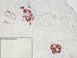
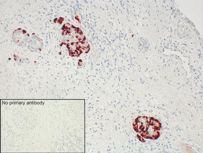
- Sections of human pancreas were dewaxed, microwaved in citric acid and incubated with Anti-GPR68 Recombinant Rabbit Monoclonal Antibody (Product # 702595, 1:500 dilution). Sections were then sequentially treated with biotinylated Anti-rabbit IgG and AB solution. Sections were then developed in AEC and lightly counterstained with hematoxylin.
Supportive validation
- Submitted by
- Invitrogen Antibodies (provider)
- Main image
- Experimental details
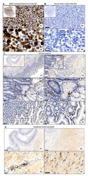
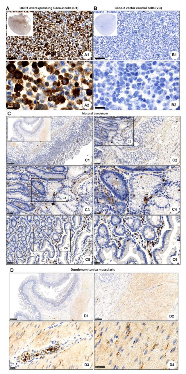
- Figure 2 Immunohistochemical detection of OGR1. ( A , B ) OGR1 (GPR68) recombinant rabbit monoclonal antibody (16H23L16) for immunohistochemical (IHC) detection of OGR1 was verified in previously validated tools. Caco-2 clone stably overexpressing OGR1 (clone U1); negative control, Caco-2 vector control (VC) cells. ( A1 , A2 ) Strong positive OGR1 staining (brown colour), predominantly in the cytoplasm, is present in OGR1-clone U1. ( B1 , B2 ) No specific staining for OGR1 in VC cells. Counterstaining with haematoxylin. Scale bar: ( A1 , B1 ) 100 um and ( A2 , B2 ) 25 um. ( C ) Localisation of OGR1 in healthy human duodenal mucosa. Scale bars: C. Insert overview image 1 mm; detail images, ( C1 ) 250 um; ( C2 ) 100 um; ( C3 , C5 ) 50 um; ( C4 , C6 ) 25 um. ( D ) Localisation of OGR1 in duodenal tunica muscularis. Scale bars: ( D1 , D2 ) 1 mm; detail images, ( D3 , D4 ) 25 um. OGR1: ovarian cancer G-protein-coupled receptor 1.
- Submitted by
- Invitrogen Antibodies (provider)
- Main image
- Experimental details
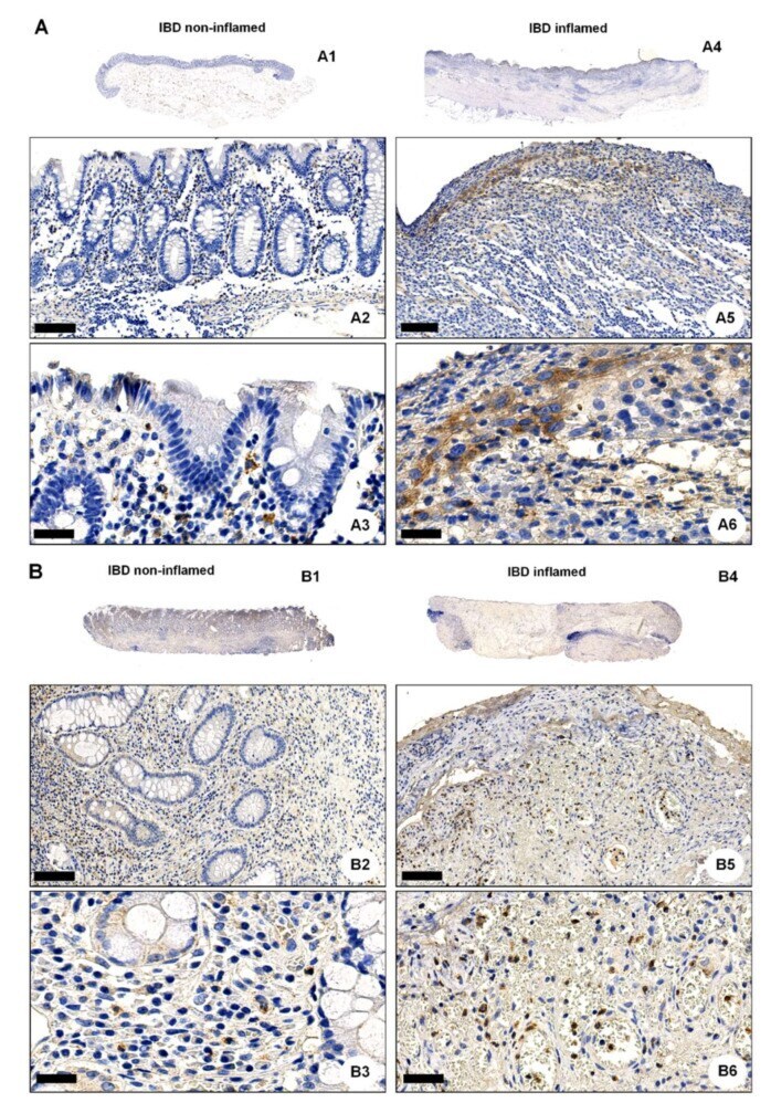
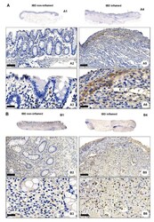
- Figure 3 OGR1 protein expression is significantly increased in the inflamed mucosa of IBD patients. Immunohistochemical detection of OGR1 using the anti-OGR1 antibody 16H23L16. Paired samples: ( A1 - A3 , B1 - B3 ) non-inflamed; ( A4 - A6 , B4 - B6 ) inflamed. Representative images shown are from two CD patients. Scale bars: ( A2 , B2 ) and ( A5 , B5 ) detail images, 100 um; ( A3 , B3 ) and ( A6 , B6 ) 25 um. ( C , D ) Representative images shown are from two non-IBD subjects. Scale bars: ( C1 , D1 ) 100 um; ( C2 , D2 ) detail images, 25 um. Intestinal resections were taken from five non-IBD subjects and paired (non-inflamed and inflamed) samples from five CD patients and five UC patients. ( E ) Total protein was isolated from colonic tissue samples and Western blotting was performed using an OGR1 (GPR68) custom mouse monoclonal antibody (AbMart). ( F ) Densitometry after normalization of OGR1 to beta-actin: five non-IBD subjects, four CD patients and four UC patients. Statistical analysis was performed using one-way ANOVA followed by Tukey's post-test (** p < 0.01; *** p < 0.001). CD: Crohn's disease; IBD: inflammatory bowel disease; OGR1: ovarian cancer G-protein-coupled receptor 1; UC: ulcerative colitis.