 Explore
 Validate
 Learn
Learn Western blot
Antibody data
- Antibody Data
- Antigen structure
- References [1]
- Comments [0]
- Validations
- Western blot [4]
- Immunocytochemistry [2]
Submit
Validation data
Reference
Comment
Report error
- Product number
- MIA1305 - Provider product page

- Provider
- Invitrogen Antibodies
- Product name
- AFP Monoclonal Antibody (P5B8)
- Antibody type
- Monoclonal
- Antigen
- Purifed from natural sources
- Description
- MIA1305 can be used for immunofluorescence analysis of AFP in the endoderm derived from human embryonic stem cells. By Western blot, MIA1305 detects endogenous AFP protein in the early hepatocyte-like cells derived from human embryonic stem cells.
- Antibody clone number
- P5B8
- Concentration
- 1.0 mg/mL
Submitted references Aglycosylated antibody-producing mice for aglycosylated antibody-lectin coupled immunoassay for the quantification of tumor markers (ALIQUAT).
Lee NE, Kim SH, Yu DY, Woo EJ, Kim MI, Seong GS, Lee SM, Ko JH, Kim YS
Communications biology 2020 Oct 30;3(1):636
Communications biology 2020 Oct 30;3(1):636
No comments: Submit comment
Supportive validation
- Submitted by
- Invitrogen Antibodies (provider)
- Main image

- Experimental details
- Western blot analysis of alpha-fetoprotein was performed by loading 10 µg of whole cell extract from H9 ESCs, H9 ESC-derived hepatoblasts, ESC-derived hepatocyte-like cells, HepG2 cells, HepaRG cells, and primary human hepatocytes in reducing conditions and 10 µL Spectra Multicolor Broad Range Protein Ladder (Product # 26634) per well onto a Novex Bolt® 4-12% Bis-Tris Plus Gel with Bolt® MOPS running buffer and Bolt® Antioxidant. Proteins were transferred to a PVDF membrane using the iBlot 2 Dry Blotting System (Product # IB21001), and blocked with 5% non-fat milk in TBST for 1 hour at room temperature. alpha-Fetoprotein was detected at 67 kDa using an alpha-fetoprotein monoclonal antibody (Product # MIA1305) at a concentration of 1 µg/mL in 5% non-fat milk in TBST overnight at 4°C on a rocking platform, followed by a goat anti-mouse secondary antibody (Product # A24518) at a dilution of 1:10,000 for 1 hour at room temperature. Chemiluminescent detection was performed using Pierce™ ECL Western Blotting Substrate (Product # 32106).

- Submitted by
- Invitrogen Antibodies (provider)
- Main image

- Experimental details
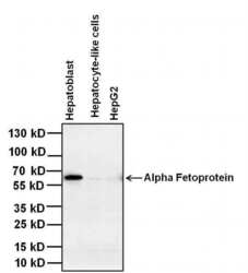
- Western blot analysis of alpha-fetoprotein was performed by loading 40 µg of the indicated whole cell lysates and 5 µL of PageRuler Plus Prestained Protein Ladder (Product # 26619) per well onto a 4-20% Tris-Glycine polyacrylamide gel (Product # WT4201BX10). Proteins were transferred to a nitrocellulose membrane using the G2 Blotter (Product # 62288), and blocked with 5% BSA in TBST for 1 hour at room temperature. Alpha-fetoprotein was detected at ~67 kDa using a alpha-fetoprotein mouse monoclonal antibody (Product # MIA1305) at a concentration of 2 µg/mL in blocking buffer overnight at 4C on a rocking platform, followed by a Goat anti-Mouse IgG (H+L) Superclonal™ Secondary Antibody, HRP conjugate (Product # A28177) at a dilution of 1:1000 for at least 30 minutes at room temperature. Chemiluminescent detection was performed using SuperSignal West Pico (Product # 34078).

- Submitted by
- Invitrogen Antibodies (provider)
- Main image
- Experimental details
- Western blot analysis of alpha-fetoprotein was performed by loading 20 µg of the indicated whole cell lysates and 5 µL of PageRuler Plus Prestained Protein Ladder (Product # 26619) per well onto a 4-20% Tris-Glycine polyacrylamide gel (Product # WT4202BX10). Proteins were transferred to a nitrocellulose membrane using the G2 Blotter (Product # 62288), and blocked with 5% BSA in TBST for 1 hour at room temperature. Alpha-fetoprotein was detected at ~67 kDa using a alpha-fetoprotein mouse monoclonal antibody (Product # MIA1305) at a concentration of 2 µg/mL in blocking buffer for 1 hour at room temperature on a rocking platform, followed by a Goat anti-Mouse IgG (H+L) Superclonal™ Secondary Antibody, HRP conjugate (Product # A28177) at a dilution of 1:1000 for at least 30 minutes at room temperature. Chemiluminescent detection was performed using SuperSignal West Pico (Product # 34078).

Supportive validation
- Submitted by
- Invitrogen Antibodies (provider)
- Main image

- Experimental details
- Immunofluorescence analysis of alpha-fetoproprotein (AFP) (green) in the endoderm derived from human ES cells. Embryoid bodies (EBs) were generated from the H9 embryonic stem cell line (WiCell Research Institute, WA09) using Gibco® KnockOut Serum Replacement. After four days in suspension culture, EBs were plated on Geltrex-coated tissue culture-treated polystyrene plates and continuously cultured for 21 days. EB cultures were then fixed and permeabilized according to the 3-Germ Layer Immunocytochemistry Kit (Product # A25538) and stained with anti-alpha-fetoprotein (Product # MIA1305, 1:200 dilution, 5 uL/mL final) at 4°C overnight. Secondary staining was completed using Alexa Fluor 488-conjugated anti-mouse IgG (Product # A-11001) and DAPI (Product # D1306) for nuclear DNA (blue) for 1 h at room temperature. Stained wells were imaged at 40X using the EVOS® FL Auto Imaging System.

- Submitted by
- Invitrogen Antibodies (provider)
- Main image

- Experimental details
- Immunofluorescent analysis of alpha-fetoprotein (green) in the endoderm derived from human ES cells. Embryoid bodies (EBs) were generated from the H9 embryonic stem cell line (WiCell Research Institute, WA09) using Gibco® KnockOut Serum Replacement. After four days in suspension culture, EBs were plated on Geltrex-coated tissue culture-treated polystyrene plates and continuously cultured for 21 days. EB cultures were then fixed and permeabilized according to the 3-Germ Layer Immunocytochemistry Kit (Product # A25538) and blocked with 3% BSA for 30 minutes at room temperature. Cells were stained with anti-alpha-fetoprotein (Product # MIA1305, 1:200 dilution, 5 µg/mL final concentration) at 4°C overnight, and then incubated with a Goat anti-Mouse IgG (H+L) Superclonal Secondary Antibody, Alexa Fluor® 488 conjugate (Product # A28175) at a dilution of 1:1000 for at least 30 minutes at a room temperature in the dark (green). Nuclei (blue) were stained with Hoechst 33342 (Product # 62249). Stained cells were imaged at 40X using the EVOS® FL Auto Imaging System.
