Explore
 Validate
 Learn
Learn Western blot
 Other assay
Antibody data
- Antibody Data
- Antigen structure
- References [4]
- Comments [0]
- Validations
- Other assay [3]
Submit
Validation data
Reference
Comment
Report error
- Product number
- PA5-21004 - Provider product page

- Provider
- Invitrogen Antibodies
- Product name
- AFP Polyclonal Antibody
- Antibody type
- Polyclonal
- Antigen
- Synthetic peptide
- Description
- A suggested positive control is human liver tissue lysate. PA5-21004 can be used with blocking peptide PEP-1118. The immunogen is located within amino acids 340 - 390 of AFP.
- Reactivity
- Human, Mouse, Rat
- Host
- Rabbit
- Isotype
- IgG
- Vial size
- 100 µg
- Concentration
- 1 mg/mL
- Storage
- Maintain refrigerated at 2-8°C for up to 3 months. For long term storage store at -20°C
Submitted references Tumor burden negatively impacts protein turnover as a proteostatic process in noncancerous liver, heart, and muscle, but not brain.
Reduced Tumorigenicity of Mouse ES Cells and the Augmented Anti-Tumor Therapeutic Effects under Parg Deficiency.
Obesity Drives STAT-1-Dependent NASH and STAT-3-Dependent HCC.
Long-term xeno-free culture of human pluripotent stem cells on hydrogels with optimal elasticity.
Brown JL, Lawrence MM, Borowik A, Oliver L, Peelor FF 3rd, Van Remmen H, Miller BF
Journal of applied physiology (Bethesda, Md. : 1985) 2021 Jul 1;131(1):72-82
Journal of applied physiology (Bethesda, Md. : 1985) 2021 Jul 1;131(1):72-82
Reduced Tumorigenicity of Mouse ES Cells and the Augmented Anti-Tumor Therapeutic Effects under Parg Deficiency.
Sonoda Y, Sasaki Y, Gunji A, Shirai H, Araki T, Imamichi S, Onodera T, Rydén AM, Watanabe M, Itami J, Honda T, Ashizawa K, Nakao K, Masutani M
Cancers 2020 Apr 24;12(4)
Cancers 2020 Apr 24;12(4)
Obesity Drives STAT-1-Dependent NASH and STAT-3-Dependent HCC.
Grohmann M, Wiede F, Dodd GT, Gurzov EN, Ooi GJ, Butt T, Rasmiena AA, Kaur S, Gulati T, Goh PK, Treloar AE, Archer S, Brown WA, Muller M, Watt MJ, Ohara O, McLean CA, Tiganis T
Cell 2018 Nov 15;175(5):1289-1306.e20
Cell 2018 Nov 15;175(5):1289-1306.e20
Long-term xeno-free culture of human pluripotent stem cells on hydrogels with optimal elasticity.
Higuchi A, Kao SH, Ling QD, Chen YM, Li HF, Alarfaj AA, Munusamy MA, Murugan K, Chang SC, Lee HC, Hsu ST, Kumar SS, Umezawa A
Scientific reports 2015 Dec 14;5:18136
Scientific reports 2015 Dec 14;5:18136
No comments: Submit comment
Supportive validation
- Submitted by
- Invitrogen Antibodies (provider)
- Main image
- Experimental details
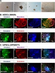
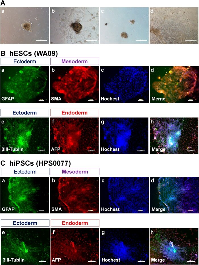
- Figure 7 Characterization of the differentiation ability of hPSCs (hESCs and hiPSCs) in vitro after culture on PVA-oligoVN hydrogels for 20 passages. ( A ) Morphology of EBs differentiated from hESCs (WA09, a,b ) and hiPSCs (HPS0077, c,d ) after culture on PVA-24h-1000 dishes under xeno-free conditions for 20 passages. ( B ) Immunostaining of an ectoderm protein ( a , GFAP; e , betaIII-tubulin), mesoderm protein ( b , SMA), and endoderm ( f , AFP) protein on hESCs (WA09) after culture on PVA-24h-1000 dishes under xeno-free conditions for 20 passages. ( c ) Hoechest staining of hESCs used in ( a,b ). ( d ) Merged picture of ( a-c ). ( g ) Hoechest staining of hESCs used in ( e , f ). (h) Merged picture of ( e-g ). The bar indicates 100 mum. ( C ) Immunostaining of an ectoderm protein ( a , GFAP; e , betaIII-tubulin), mesoderm protein ( b , SMA), and endoderm ( f , AFP) protein on hiPSCs (HPS0077) after culture on PVA-24h-1000 dishes under xeno-free conditions for 20 passages. ( c ) Hoechest staining of hESCs used in ( a,b ). ( d ) Merged picture of ( a-c ). ( g ) Hoechest staining of hESCs used in ( e , f ). ( h ) Merged picture of ( e-g ). The bar indicates 100 mum.
- Submitted by
- Invitrogen Antibodies (provider)
- Main image

- Experimental details
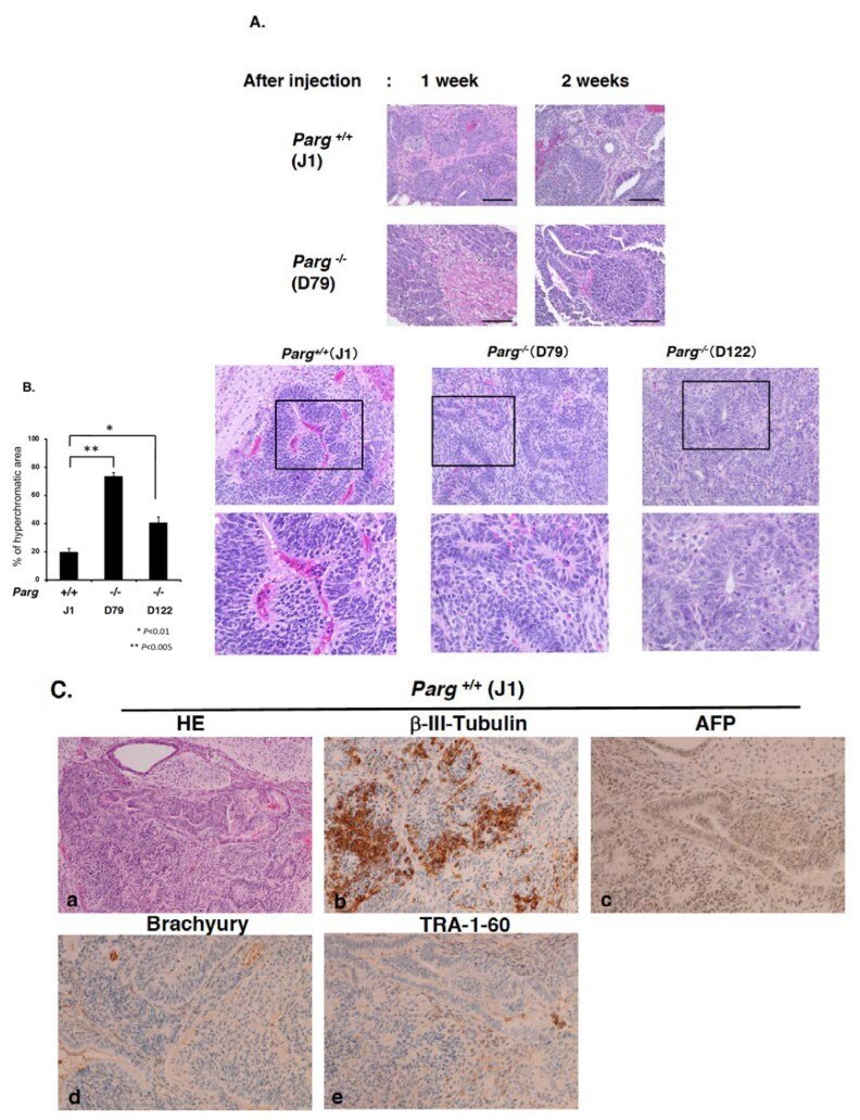
- Figure 2 Hematoxylin-eosin staining of tumor tissues derived from mouse ES cells. ( A ) Hematoxylin-eosin staining of tumor tissues derived from mouse ES cells 1 and 2 weeks after injection. Upper panels, wild-type J1. Lower panels, Parg -/- . ( B ) The left graph shows the percentage of hyperchromatic area of the tumors 4 weeks after injection. Percentage of hematoxylin-positive hyperchromatic area in the total area of tumor section was measured for each tumor. * p < 0.01, ** p < 0.005. Right panels show the typical hyperchromatic areas of hematoxylin-eosin staining of tumors 4 weeks after injection. Upper panels, x20 magnification (Squares show magnified regions in the lower panels. Lower panels, x40 magnification. The tumors showed heterogeneous cell components containing primitive neuroepithelial components and embryonal carcinoma components. ( C ) HE staining and immunostaining of the tumors at 4 weeks with antibodies against b-III-tubulin, ectoderm marker; AFP, endoderm marker (x20 magnification). Hematoxylin-eosin staining, x10 magnification. The mixed staining pattern of ectodermal and endodermal markers was observed in hyperchromatic regions of Parg -/- tumors at 4 weeks. ( D ) Immunostaining of the tumors at 4 weeks after injection with antibody against anti-PAR. Right panels in D are magnified images, Bars, 50 mm (left panels in D), 20 mm (right panels in D). PAR staining was observed occasionally in the cell nuclei in the Parg -/- tumor but not in the Parg +/+ tum
- Submitted by
- Invitrogen Antibodies (provider)
- Main image
- Experimental details
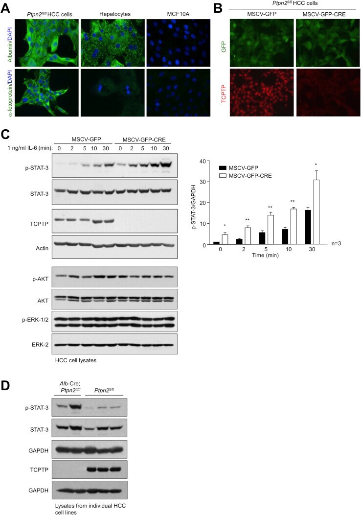
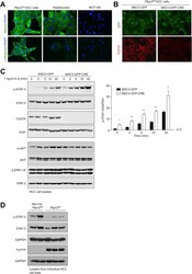
- Figure S7 TCPTP Deletion in HCC Cells Promotes STAT3 Signaling, Related to Figure 7 (A-C) Tumors from DEN-treated Ptpn2 fl/fl mice were dissociated and HCC cell lines established. (A) Ptpn2 fl/fl HCC cells, primary hepatocytes from chow-fed C57BL/6 mice and MCF10A immortalized mammary epithelial cells were processed for immunofluorescence microscopy monitoring for the presence of the hepatocyte marker albumin, or the liver cancer marker alpha-fetoprotein. Ptpn2 fl/fl HCC cells were transduced with retroviruses encoding GFP alone (MSCV-GFP) or GFP and Cre recombinase (MSCV-GFP-CRE) and sorted twice for GFP and either processed for (B) immunofluorescence microscopy monitoring for GFP and TCPTP, or (C) serum starved and stimulated with IL-6 (1 ng/ml) and then processed for immunoblotting monitoring for STAT-3 signaling. Quantified results (means +- SEM) are shown for the indicated number of experiments with significance determined using a Student's t test. (D) Tumors from DEN-treated Ptpn2 fl/fl or Alb -Cre; Ptpn2 fl/fl were dissociated and HCC cell lines established. HCC cells were serum starved and processed for immunoblotting.