 Explore
 Validate
 Learn
Learn Immunocytochemistry
Antibody data
- Antibody Data
- Antigen structure
- References [1]
- Comments [0]
- Validations
- Immunocytochemistry [3]
Submit
Validation data
Reference
Comment
Report error
- Product number
- 53-6583-82 - Provider product page

- Provider
- Invitrogen Antibodies
- Product name
- alpha-Fetoprotein Monoclonal Antibody (AFP3), Alexa Fluor™ 488, eBioscience™
- Antibody type
- Monoclonal
- Antigen
- Other
- Description
- Description: This AFP3 monoclonal antibody reacts with human alpha-fetoprotein (AFP). This 70-kDa secretory protein is a member of the albumin gene family. Synthesized by the yolk sac and fetal liver during embryogenesis, AFP protein levels are highest in fetal serum. After birth, serum AFP levels decrease dramatically. In fact, AFP is nearly undetectable in normal adult serum. However, hepatocellular carcinoma and germ cell teratoblastoma, as well as liver regeneration, viral hepatitis, and cirrhosis, leads to elevated AFP serum levels in adults. As such, detection of this protein is frequently used as a diagnostic marker for these conditions. When performing western blotting or immunohistochemistry on paraffin section, we recommend the use of monoclonal antibody 1E8 (Product # 14-9499). Applications Reported: This AFP3 antibody has been reported for use in immunocytochemistry. Applications Tested: This AFP3 antibody has been tested by immunofluorescent staining of formaldehyde-fixed and permeabilized HepG2 cells. This can be used at less than or equal to 10 µg/mL. It is recommended that the antibody be titrated for optimal performance in the assay of interest. Excitation: 488 nm; Emission: 519 nm; Laser: Blue Laser. Filtration: 0.2 µm post-manufacturing filtered.
- Reactivity
- Human
- Host
- Mouse
- Conjugate
- Green dye
- Isotype
- IgG
- Antibody clone number
- AFP3
- Vial size
- 100 µg
- Concentration
- 0.5 mg/mL
- Storage
- 4° C, store in dark, DO NOT FREEZE!
Submitted references Generation of monoclonal antibodies to alpha-fetoprotein and application in solid-phase enzyme immunoassay.
Kuo CY, Fu J, Yeh MY, Su SL, Lee CY
Biotechnology and applied biochemistry 1989 Feb;11(1):96-104
Biotechnology and applied biochemistry 1989 Feb;11(1):96-104
No comments: Submit comment
Supportive validation
- Submitted by
- Invitrogen Antibodies (provider)
- Main image

- Experimental details
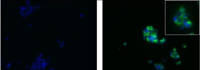
- Immunocytochemistry of fixed and permeabilized HepG2 cells using 10 µg/mL of Mouse IgG1 Isotype Control Alexa Fluor® 488 (Product # 53-4714-42) (left) or Anti-Human AFP Alexa Fluor® 488 (right, high magnification shown in inset). Nuclei were counterstained with DAPI.
- Conjugate
- Green dye
- Submitted by
- Invitrogen Antibodies (provider)
- Main image
- Experimental details
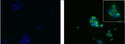
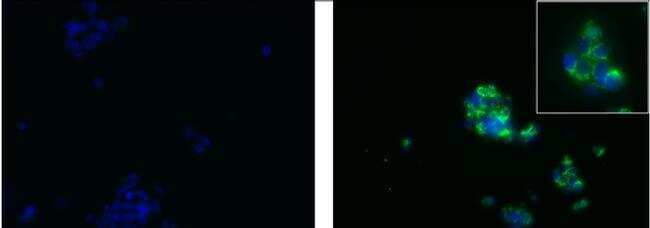
- Immunocytochemistry of fixed and permeabilized HepG2 cells using 10 µg/mL of Mouse IgG1 Isotype Control Alexa Fluor® 488 (Product # 53-4714-42) (left) or Anti-Human AFP Alexa Fluor® 488 (right, high magnification shown in inset). Nuclei were counterstained with DAPI.
- Conjugate
- Green dye
- Submitted by
- Invitrogen Antibodies (provider)
- Main image

- Experimental details
- Immunofluorescence analysis of AFP was performed using 70% confluent log phase Hep G2 cells. The cells were fixed with 4% paraformaldehyde for 10 minutes, permeabilized with 0.1% Triton™ X-100 for 15 minutes, and blocked with 1% BSA for 1 hour at room temperature. The cells were labeled with AFP Mouse Monoclonal Antibody (Product # 53-6583-82) at 5 µg/mL in 0.1% BSA, incubated at 4 degree celsius overnight (Panel a: green). Nuclei (Panel b: blue) were stained with ProLong™ Diamond Antifade Mountant with DAPI (Product # P36962). F-actin (Panel c: red) was stained with Rhodamine Phalloidin (Product # R415, 1:300). Panel d represents the merged image showing nuclear localization. Panel e shows AFP negative cell line HeLa with no signal. Panel f represents control cells with Isotype control to assess background. The images were captured at 60X magnification.
- Conjugate
- Green dye
