 Explore
 Validate
 Learn
Learn Western blot
 ELISA
ELISA Immunocytochemistry
Antibody data
- Antibody Data
- Antigen structure
- References [0]
- Comments [0]
- Validations
- Immunocytochemistry [3]
Submit
Validation data
Reference
Comment
Report error
- Product number
- MA5-14665 - Provider product page

- Provider
- Invitrogen Antibodies
- Product name
- AFP Monoclonal Antibody (F1-6P2A8-P2B9A9)
- Antibody type
- Monoclonal
- Antigen
- Purifed from natural sources
- Description
- MA5-14665 can be used for immunofluorescent analysis of AFP in the endoderm derived from human embryonic stem cells. By Western blot, MA5-14665 detects endogenous AFP protein in the early hepatocyte-like cells derived from human embryonic stem cells. Product MA514665 is a smaller package size of MIA1301 (formerly sold as a Seradyn product).
- Reactivity
- Human
- Host
- Mouse
- Isotype
- IgG
- Antibody clone number
- F1-6P2A8-P2B9A9
- Vial size
- 100 μg
- Concentration
- 1 mg/mL
- Storage
- Maintain refrigerated at 2-8°C for up to 6 months. For long term storage store at -20°C
No comments: Submit comment
Supportive validation
- Submitted by
- Invitrogen Antibodies (provider)
- Main image
- Experimental details
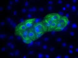
- Immunofluorescence analysis of alpha-fetoproprotein (AFP) (green) in the endoderm derived from human ES cells. Embryoid bodies (EBs) were generated from the H9 embryonic stem cell line (WiCell Research Institute, WA09) using Gibco® KnockOut Serum Replacement. After four days in suspension culture, EBs were plated on Geltrex-coated tissue culture-treated polystyrene plates and continuously cultured for 21 days. EB cultures were then fixed and permeabilized according to the 3-Germ Layer Immunocytochemistry Kit (Product # A25538) and stained with anti-alpha-fetoprotein (Product # MA5-14665, 1:200 dilution, 5 uL/mL final) at 4°C overnight. Secondary staining was completed using Alexa Fluor 488-conjugated anti-mouse IgG (Product # A-11001) and DAPI (Product # D1306) for nuclear DNA (blue) for 1 h at room temperature. Stained wells were imaged at 40X using the EVOS® FL Auto Imaging System.

- Submitted by
- Invitrogen Antibodies (provider)
- Main image
- Experimental details
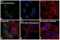
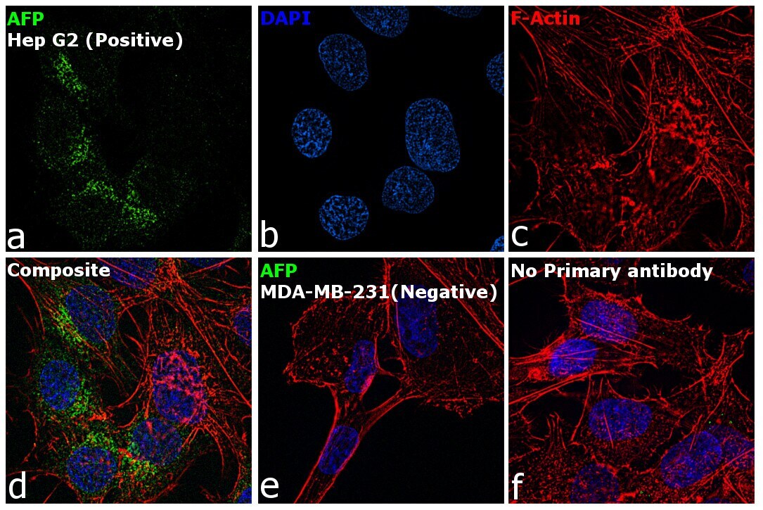
- Immunofluorescence analysis of AFP was performed using 70% confluent log phase Hep G2 and MDA-MB-231 cells. The cells were fixed with 4% paraformaldehyde for 10 minutes, permeabilized with 0.1% Triton™ X-100 for 15 minutes, and blocked with 2% BSA for 1 hour at room temperature. The cells were labeled with AFP Mouse Monoclonal Antibody (Product # MA5-14665) at 5 µg/mL in 0.1% BSA and incubated overnight at 4 degree and then labeled with Goat anti-Mouse IgG (H+L) Superclonal™ Recombinant Secondary Antibody, Alexa Fluor® 488 (Product # A28175) at a dilution of 1:2000 for 45 minutes at room temperature (Panel a: green). Nuclei (Panel b: blue) were stained with ProLong™ Diamond Antifade Mountant with DAPI (Product # P36962). F-actin (Panel c: red) was stained with Rhodamine Phalloidin (Product # R415, 1:300). Panel d represents the composite image showing cytoplasmic and golgi staining of AFP in Hep G2. Panel e represents the merged image of MDA-MB-231 cells which do not have AFP expression. Panel f represents control cells with no primary antibody to assess background. The images were captured at 60X magnification..
- Submitted by
- Invitrogen Antibodies (provider)
- Main image

- Experimental details
- Immunofluorescence analysis of AFP was performed using 70% confluent log phase Hep G2 and MDA-MB-231 cells. The cells were fixed with 4% paraformaldehyde for 10 minutes, permeabilized with 0.1% Triton™ X-100 for 15 minutes, and blocked with 2% BSA for 1 hour at room temperature. The cells were labeled with AFP Mouse Monoclonal Antibody (Product # MA5-14665) at 5 µg/mL in 0.1% BSA and incubated overnight at 4 degree and then labeled with Goat anti-Mouse IgG (H+L) Superclonal™ Recombinant Secondary Antibody, Alexa Fluor® 488 (Product # A28175) at a dilution of 1:2000 for 45 minutes at room temperature (Panel a: green). Nuclei (Panel b: blue) were stained with ProLong™ Diamond Antifade Mountant with DAPI (Product # P36962). F-actin (Panel c: red) was stained with Rhodamine Phalloidin (Product # R415, 1:300). Panel d represents the composite image showing cytoplasmic and golgi staining of AFP in Hep G2. Panel e represents the merged image of MDA-MB-231 cells which do not have AFP expression. Panel f represents control cells with no primary antibody to assess background. The images were captured at 60X magnification..
