Explore
 Validate
 Learn
Learn Western blot
Antibody data
- Antibody Data
- Antigen structure
- References [25]
- Comments [0]
- Validations
- Western blot [3]
- Immunocytochemistry [1]
- Flow cytometry [2]
Submit
Validation data
Reference
Comment
Report error
- Product number
- MAB1368 - Provider product page

- Provider
- R&D Systems
- Product name
- Human/Mouse alpha-Fetoprotein/AFP Antibody
- Antibody type
- Monoclonal
- Description
- Protein A or G purified from hybridoma culture supernatant. Detects human alpha-Fetoprotein/AFP in direct ELISAs and Western blots.
- Reactivity
- Human, Mouse
- Host
- Mouse
- Conjugate
- Unconjugated
- Isotype
- IgG
- Antibody clone number
- 189502
- Vial size
- 100 ug
- Concentration
- LYOPH
- Storage
- Use a manual defrost freezer and avoid repeated freeze-thaw cycles. 12 months from date of receipt, -20 to -70 °C as supplied. 1 month, 2 to 8 °C under sterile conditions after reconstitution. 6 months, -20 to -70 °C under sterile conditions after reconstitution.
Submitted references Chromosome Transplantation: Correction of the Chronic Granulomatous Disease Defect in Mouse Induced Pluripotent Stem Cells.
Generation and characterization of the human iPSC line CABi001-A from a patient with retinitis pigmentosa caused by a novel mutation in PRPF31 gene.
NEK1 loss-of-function mutation induces DNA damage accumulation in ALS patient-derived motoneurons.
The oncogene Etv5 promotes MET in somatic reprogramming and orchestrates epiblast/primitive endoderm specification during mESCs differentiation.
Generation of nine induced pluripotent stem cell lines as an ethnic diversity panel.
Generation of a human iPSC line from a patient with congenital glaucoma caused by mutation in CYP1B1 gene.
Generation of D1-1 TALEN isogenic control cell line from Dravet syndrome patient iPSCs using TALEN-mediated editing of the SCN1A gene.
Generation of a gene-corrected isogenic control iPSC line from cystic fibrosis patient-specific iPSCs homozygous for p.Phe508del mutation mediated by TALENs and ssODN.
One Year Follow-Up Risk Assessment in SKH-1 Mice and Wounds Treated with an Argon Plasma Jet.
Altered Biomarkers in Trophoblast Cells Obtained Noninvasively Prior to Clinical Manifestation of Perinatal Disease.
Generation of Naïve Bovine Induced Pluripotent Stem Cells Using PiggyBac Transposition of Doxycycline-Inducible Transcription Factors.
Controlled Growth and the Maintenance of Human Pluripotent Stem Cells by Cultivation with Defined Medium on Extracellular Matrix-Coated Micropatterned Dishes.
Generation of human induced pluripotent stem (Ips) cells in serum- and feeder-free defined culture and TGF-Β1 regulation of pluripotency.
Cell-autonomous correction of ring chromosomes in human induced pluripotent stem cells.
Lin28b is sufficient to drive liver cancer and necessary for its maintenance in murine models.
Enzyme-free passage of human pluripotent stem cells by controlling divalent cations.
Identification of transcription factors for lineage-specific ESC differentiation.
Establishment of induced pluripotent stem cells from centenarians for neurodegenerative disease research.
Differentiation and transplantation of functional pancreatic beta cells generated from induced pluripotent stem cells derived from a type 1 diabetes mouse model.
Small RNA-mediated regulation of iPS cell generation.
Induction of pluripotent stem cells from human third molar mesenchymal stromal cells.
Effective generation of iPS cells from CD34+ cord blood cells by inhibition of p53.
Expansion of CD133-expressing liver cancer stem cells in liver-specific phosphatase and tensin homolog deleted on chromosome 10-deleted mice.
Induction of pluripotent stem cells from adult human fibroblasts by defined factors.
Wnt/beta-catenin/CBP signaling maintains long-term murine embryonic stem cell pluripotency.
Castelli A, Susani L, Menale C, Muggeo S, Caldana E, Strina D, Cassani B, Recordati C, Scanziani E, Ficara F, Villa A, Vezzoni P, Paulis M
Stem cells (Dayton, Ohio) 2019 Jul;37(7):876-887
Stem cells (Dayton, Ohio) 2019 Jul;37(7):876-887
Generation and characterization of the human iPSC line CABi001-A from a patient with retinitis pigmentosa caused by a novel mutation in PRPF31 gene.
de la Cerda B, Díez-Lloret A, Ponte B, Vallés-Saiz L, Calado SM, Rodríguez-Bocanegra E, Garcia-Delgado AB, Moya-Molina M, Bhattacharya SS, Díaz-Corrales FJ
Stem cell research 2019 Apr;36:101426
Stem cell research 2019 Apr;36:101426
NEK1 loss-of-function mutation induces DNA damage accumulation in ALS patient-derived motoneurons.
Higelin J, Catanese A, Semelink-Sedlacek LL, Oeztuerk S, Lutz AK, Bausinger J, Barbi G, Speit G, Andersen PM, Ludolph AC, Demestre M, Boeckers TM
Stem cell research 2018 Jul;30:150-162
Stem cell research 2018 Jul;30:150-162
The oncogene Etv5 promotes MET in somatic reprogramming and orchestrates epiblast/primitive endoderm specification during mESCs differentiation.
Zhang J, Cao H, Xie J, Fan C, Xie Y, He X, Liao M, Zhang S, Wang H
Cell death & disease 2018 Feb 14;9(2):224
Cell death & disease 2018 Feb 14;9(2):224
Generation of nine induced pluripotent stem cell lines as an ethnic diversity panel.
Gao X, Yourick JJ, Sprando RL
Stem cell research 2018 Aug;31:193-196
Stem cell research 2018 Aug;31:193-196
Generation of a human iPSC line from a patient with congenital glaucoma caused by mutation in CYP1B1 gene.
Bolinches-Amorós A, Lukovic D, Castro AA, León M, Kamenarova K, Kaneva R, Jendelova P, Blanco-Kelly F, Ayuso C, Cortón M, Erceg S
Stem cell research 2018 Apr;28:96-99
Stem cell research 2018 Apr;28:96-99
Generation of D1-1 TALEN isogenic control cell line from Dravet syndrome patient iPSCs using TALEN-mediated editing of the SCN1A gene.
Tanaka Y, Sone T, Higurashi N, Sakuma T, Suzuki S, Ishikawa M, Yamamoto T, Mitsui J, Tsuji H, Okano H, Hirose S
Stem cell research 2018 Apr;28:100-104
Stem cell research 2018 Apr;28:100-104
Generation of a gene-corrected isogenic control iPSC line from cystic fibrosis patient-specific iPSCs homozygous for p.Phe508del mutation mediated by TALENs and ssODN.
Merkert S, Bednarski C, Göhring G, Cathomen T, Martin U
Stem cell research 2017 Aug;23:95-97
Stem cell research 2017 Aug;23:95-97
One Year Follow-Up Risk Assessment in SKH-1 Mice and Wounds Treated with an Argon Plasma Jet.
Schmidt A, Woedtke TV, Stenzel J, Lindner T, Polei S, Vollmar B, Bekeschus S
International journal of molecular sciences 2017 Apr 19;18(4)
International journal of molecular sciences 2017 Apr 19;18(4)
Altered Biomarkers in Trophoblast Cells Obtained Noninvasively Prior to Clinical Manifestation of Perinatal Disease.
Bolnick JM, Kohan-Ghadr HR, Fritz R, Bolnick AD, Kilburn BA, Diamond MP, Armant DR, Drewlo S
Scientific reports 2016 Sep 23;6:32382
Scientific reports 2016 Sep 23;6:32382
Generation of Naïve Bovine Induced Pluripotent Stem Cells Using PiggyBac Transposition of Doxycycline-Inducible Transcription Factors.
Kawaguchi T, Tsukiyama T, Kimura K, Matsuyama S, Minami N, Yamada M, Imai H
PloS one 2015;10(8):e0135403
PloS one 2015;10(8):e0135403
Controlled Growth and the Maintenance of Human Pluripotent Stem Cells by Cultivation with Defined Medium on Extracellular Matrix-Coated Micropatterned Dishes.
Takenaka C, Miyajima H, Yoda Y, Imazato H, Yamamoto T, Gomi S, Ohshima Y, Kagawa K, Sasaki T, Kawamata S
PloS one 2015;10(6):e0129855
PloS one 2015;10(6):e0129855
Generation of human induced pluripotent stem (Ips) cells in serum- and feeder-free defined culture and TGF-Β1 regulation of pluripotency.
Yamasaki S, Taguchi Y, Shimamoto A, Mukasa H, Tahara H, Okamoto T
PloS one 2014;9(1):e87151
PloS one 2014;9(1):e87151
Cell-autonomous correction of ring chromosomes in human induced pluripotent stem cells.
Bershteyn M, Hayashi Y, Desachy G, Hsiao EC, Sami S, Tsang KM, Weiss LA, Kriegstein AR, Yamanaka S, Wynshaw-Boris A
Nature 2014 Mar 6;507(7490):99-103
Nature 2014 Mar 6;507(7490):99-103
Lin28b is sufficient to drive liver cancer and necessary for its maintenance in murine models.
Nguyen LH, Robinton DA, Seligson MT, Wu L, Li L, Rakheja D, Comerford SA, Ramezani S, Sun X, Parikh MS, Yang EH, Powers JT, Shinoda G, Shah SP, Hammer RE, Daley GQ, Zhu H
Cancer cell 2014 Aug 11;26(2):248-61
Cancer cell 2014 Aug 11;26(2):248-61
Enzyme-free passage of human pluripotent stem cells by controlling divalent cations.
Ohnuma K, Fujiki A, Yanagihara K, Tachikawa S, Hayashi Y, Ito Y, Onuma Y, Chan T, Michiue T, Furue MK, Asashima M
Scientific reports 2014 Apr 11;4:4646
Scientific reports 2014 Apr 11;4:4646
Identification of transcription factors for lineage-specific ESC differentiation.
Yamamizu K, Piao Y, Sharov AA, Zsiros V, Yu H, Nakazawa K, Schlessinger D, Ko MS
Stem cell reports 2013;1(6):545-59
Stem cell reports 2013;1(6):545-59
Establishment of induced pluripotent stem cells from centenarians for neurodegenerative disease research.
Yagi T, Kosakai A, Ito D, Okada Y, Akamatsu W, Nihei Y, Nabetani A, Ishikawa F, Arai Y, Hirose N, Okano H, Suzuki N
PloS one 2012;7(7):e41572
PloS one 2012;7(7):e41572
Differentiation and transplantation of functional pancreatic beta cells generated from induced pluripotent stem cells derived from a type 1 diabetes mouse model.
Jeon K, Lim H, Kim JH, Thuan NV, Park SH, Lim YM, Choi HY, Lee ER, Kim JH, Lee MS, Cho SG
Stem cells and development 2012 Sep 20;21(14):2642-55
Stem cells and development 2012 Sep 20;21(14):2642-55
Small RNA-mediated regulation of iPS cell generation.
Li Z, Yang CS, Nakashima K, Rana TM
The EMBO journal 2011 Mar 2;30(5):823-34
The EMBO journal 2011 Mar 2;30(5):823-34
Induction of pluripotent stem cells from human third molar mesenchymal stromal cells.
Oda Y, Yoshimura Y, Ohnishi H, Tadokoro M, Katsube Y, Sasao M, Kubo Y, Hattori K, Saito S, Horimoto K, Yuba S, Ohgushi H
The Journal of biological chemistry 2010 Sep 17;285(38):29270-8
The Journal of biological chemistry 2010 Sep 17;285(38):29270-8
Effective generation of iPS cells from CD34+ cord blood cells by inhibition of p53.
Takenaka C, Nishishita N, Takada N, Jakt LM, Kawamata S
Experimental hematology 2010 Feb;38(2):154-62
Experimental hematology 2010 Feb;38(2):154-62
Expansion of CD133-expressing liver cancer stem cells in liver-specific phosphatase and tensin homolog deleted on chromosome 10-deleted mice.
Rountree CB, Ding W, He L, Stiles B
Stem cells (Dayton, Ohio) 2009 Feb;27(2):290-9
Stem cells (Dayton, Ohio) 2009 Feb;27(2):290-9
Induction of pluripotent stem cells from adult human fibroblasts by defined factors.
Takahashi K, Tanabe K, Ohnuki M, Narita M, Ichisaka T, Tomoda K, Yamanaka S
Cell 2007 Nov 30;131(5):861-72
Cell 2007 Nov 30;131(5):861-72
Wnt/beta-catenin/CBP signaling maintains long-term murine embryonic stem cell pluripotency.
Miyabayashi T, Teo JL, Yamamoto M, McMillan M, Nguyen C, Kahn M
Proceedings of the National Academy of Sciences of the United States of America 2007 Mar 27;104(13):5668-73
Proceedings of the National Academy of Sciences of the United States of America 2007 Mar 27;104(13):5668-73
No comments: Submit comment
Supportive validation
- Submitted by
- R&D Systems (provider)
- Main image
- Experimental details
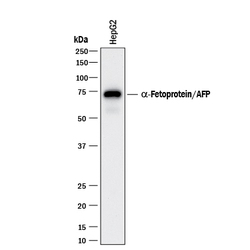
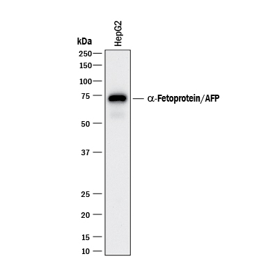
- Detection of alpha-Fetoprotein/AFP by Western Blot. Western blot shows lysates of HepG2 human hepatocellular carcinoma cell line. PVDF membrane was probed with 0.5 µg/mL of Mouse Anti-Human/Mouse alpha-Fetoprotein/AFP Monoclonal Antibody (Catalog # MAB1368) followed by HRP-conjugated Anti-Mouse IgG Secondary Antibody (Catalog # HAF018). A specific band was detected for alpha-Fetoprotein/AFP at approximately 70 kDa (as indicated). This experiment was conducted under reducing conditions and using Immunoblot Buffer Group 1.
- Submitted by
- R&D Systems (provider)
- Main image
- Experimental details
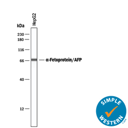
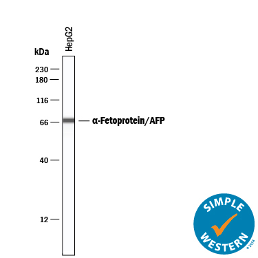
- Detection of Human alpha-Fetoprotein/AFP by Simple WesternTM. Simple Western lane view shows lysates of HepG2 human hepatocellular carcinoma cell line, loaded at 0.2 mg/mL. A specific band was detected for alpha-Fetoprotein/AFP at approximately 70 kDa (as indicated) using 5 µg/mL of Mouse Anti-Human/Mouse alpha-Fetoprotein/AFP Monoclonal Antibody (Catalog # MAB1368). This experiment was conducted under reducing conditions and using the 12-230 kDa separation system.
- Submitted by
- R&D Systems (provider)
- Main image
- Experimental details
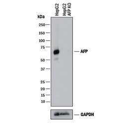
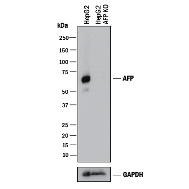
- Western Blot Shows Human alpha-Fetoprotein/AFP Specificity by Using Knockout Cell Line. Western blot shows lysates of HepG2 human hepatocellular carcinoma parental cell line and alpha-Fetoprotein/AFP knockout HepG2 cell line (KO). PVDF membrane was probed with 0.1 µg/mL of Mouse Anti-Human/Mouse alpha-Fetoprotein/AFP Monoclonal Antibody (Catalog # MAB1368) followed by HRP-conjugated Anti-Mouse IgG Secondary Antibody (Catalog # HAF018). A specific band was detected for alpha-Fetoprotein/AFP at approximately 70 kDa (as indicated) in the parental HeLa cell line, but is not detectable in knockout HeLa cell line. GAPDH (Catalog # AF5718) is shown as a loading control. This experiment was conducted under reducing conditions and using Immunoblot Buffer Group 1.
Supportive validation
- Submitted by
- R&D Systems (provider)
- Main image
- Experimental details
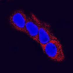
- alpha-Fetoprotein/AFP in HepG2 Human Cell Line. alpha-Fetoprotein/AFP was detected in immersion fixed HepG2 human hepatocellular carcinoma cell line using Mouse Anti-Human/Mouse alpha-Fetoprotein/AFP Monoclonal Antibody (Catalog # MAB1368) at 25 µg/mL for 3 hours at room temperature. Cells were stained using the NorthernLights™ 557-conjugated Anti-Mouse IgG Secondary Antibody (red; Catalog # NL007) and counterstained with DAPI (blue). Specific staining was localized to cytoplasm. View our protocol for Fluorescent ICC Staining of Cells on Coverslips.

Supportive validation
- Submitted by
- R&D Systems (provider)
- Main image
- Experimental details
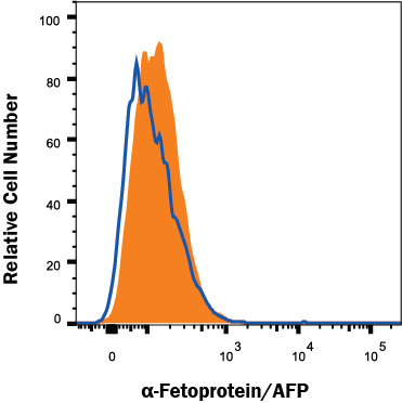
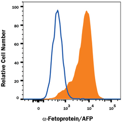
- Detection of alpha-Fetoprotein/AFP in HepG2 Human Cell Line by Flow Cytometry. HepG2 human hepatocellular carcinoma cell line was stained with Mouse Anti-Human/Mouse alpha-Fetoprotein/AFP Monoclonal Antibody (Catalog # MAB1368, filled histogram) or isotype control antibody (Catalog # MAB002, open histogram), followed by Phycoerythrin-conjugated Anti-Mouse IgG Secondary Antibody (Catalog # F0102B). To facilitate intracellular staining, cells were fixed with Flow Cytometry Fixation Buffer (Catalog # FC004) and permeabilized with Flow Cytometry Permeabilization/Wash Buffer I (Catalog # FC005). View our protocol for Staining Intracellular Molecules.
- Submitted by
- R&D Systems (provider)
- Main image

- Experimental details
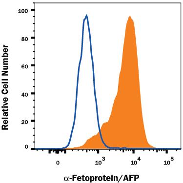
- alpha-Fetoprotein/AFP Specificity is Shown by Flow Cytometry in Knockout Cell Line. alpha-Fetoprotein/AFP knockout HepG2 hepatocellular carcinoma cell line was stained with Mouse Anti-Human alpha-Fetoprotein/AFP Monoclonal Antibody (Catalog # MAB1368, filled histogram) or isotype control antibody (Catalog # MAB002, open histogram) followed by PE-conjugated Goat anti-Mouse IgG Secondary Antibody (Catalog # F0102B). No staining in the alpha-Fetoprotein/AFP knockout HepG2 cell line was observed. View our protocol for Staining Membrane-associated Proteins.